Lesson 1: Clinical Disorders – Part D
PART D
Sleep is a Dynamic Process
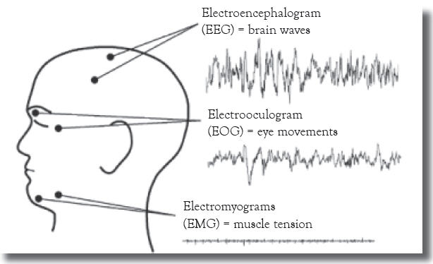
Sleep is not a passive event, but rather an active process involving characteristic physiological changes in the organs of the body. Scientists study sleep by measuring the electrical changes in the brain using electroencephalograms (EEGs). Typically, electrodes are placed on the scalp in a symmetrical pattern. The electrodes measure very small voltages that scientists think are caused by synchronized activity in very large numbers of synapses (nerve connections) in the brain’s outer layers (cerebral cortex). EEG data are represented by curves that are classified according to their frequencies. The wavy lines of the EEG are called brain waves. An electrooculogram (EOG) uses electrodes on the skin near the eye to measure changes in voltage as the eye rotates in its socket. Scientists also measure the electrical activity associated with active muscles by using electromyograms (EMGs). In this technique, electrodes are placed on the skin overlaying a muscle. In humans, the electrodes are placed under the chin because muscles in this area demonstrate very dramatic changes during the various stages of sleep.
In practice, EEGs, EOGs, and EMGs are recorded simultaneously on continuously moving chart paper or digitized by a computer and displayed on a high-resolution monitor.
This allows the relationships among the three measurements to be seen immediately. The patterns of activity in these three systems provide the basis for classifying the different types of sleep.
Figure 3.1:
Placement of electrodes to determine EEG, EOG, and EMG
Studying these events has led to the identification of two basic stages, or states, of sleep: non–rapid eye movement (NREM) and rapid eye movement (REM). Sleep is a highly organized sequence of events that follows a regular, cyclic program each night. Thus, the EEG, EMG, and EOG patterns change in predictable ways several times during a single sleep period. NREM sleep is divided into four stages according to the amplitude and frequency of brain wave activity. In general, the EEG pattern of NREM sleep is slower, often more regular, and usually of higher voltage than wakefulness. As sleep gets deeper, the brain waves get slower and have greater amplitude. NREM Stage 1 is very light sleep; NREM Stage 2 has special brain waves called sleep spindles and K complexes; NREM Stages 3 and 4 show increasingly more high-voltage slow waves. In NREM Stage 4, it is extremely difficult to be awakened by external stimuli. The muscle activity of NREM sleep is low, but the muscles retain their ability to function. Eye movements normally do not occur during NREM sleep, except for very slow eye movements, usually at the beginning. The body’s general physiology during these stages is fairly similar to the wake state.
Figure 3.2: Characteristic EEG, EOG, and EMG patterns for wakefulness, REM sleep, and NREM sleep. Each of the nine patterns was made over a period of about three seconds [a three-second sample].
Figure 3.2:
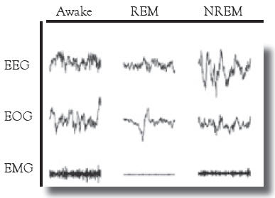
The EEG recorded during REM sleep shows very fast and desynchronized activity that is more random than that recorded during NREM sleep. It actually looks similar to the EEG (low voltage with a faster mix of frequencies) from when we are awake. REM sleep is characterized by bursts of rapid eye movements. The eyes are not constantly moving, but they dart back and forth or up and down. They also stop for a while and then jerk back and forth again. Always, and just like waking eye movements, both eyes move together in the same direction. Some scientists believe that the eye movements of REM sleep relate to the visual images of dreams, but why they exist and what function they serve, if any, remain unknown. Additionally, while muscle tone is normal in NREM sleep, we are almost completely paralyzed in REM sleep. Although the muscles that move our bodies go limp, other important muscles continue to function in REM sleep. These include the heart, diaphragm, eye muscles, and smooth muscles such as those of the intestines and blood vessels. The paralysis of muscles in the arms and legs and under the chin show electrical silence in REM sleep. On an EMG, the recording produces a flat line. Small twitches can break through this paralysis and look like tiny blips on the flat line.