Module 6 Intro
1. Module 6 Intro
1.6. Page 4
Module 6—The Motor System and Homeostasis
Muscle Hierarchy—The Way That Muscles Are Organized
Muscle tissue is organized in a hierarchical manner.

Helene Colbourne et al, Inquiry into Biology (Toronto: McGraw-Hill Ryerson, 2007), p. 335. Reproduced by permission.
muscle fibre: a single muscle cell
actin: a thin myofilament consisting of two strands of actin protein molecules wrapped around each other
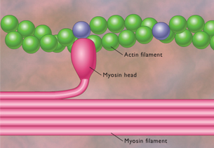
Actin works with myosin to produce muscle contractions.
myosin: a thick myofilament consisting of two strands of myosin molecules wound around each other
One end consists of a long rod, while the other end consists of a double-headed globular region. Myosin works with actin to produce muscle contractions.
Have you ever cut up raw meat in preparation for cooking? If you haven’t thought about it before, the meat that you eat is muscle. The translucent covering of a chicken breast that fat is often attached to is connective tissue, or tendon. A chicken breast, if examined closely with a microscope, would display groups of muscle fibre bundles. Since a chicken breast would be considered a skeletal muscle, muscle cells would show striations under the microscope. Essentially, when you eat meat, you are consuming the contractile proteins actin and myosin.
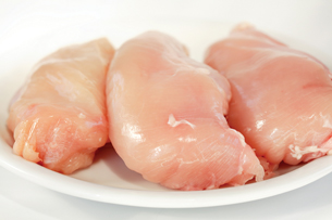
© Ingrid Balabanova/shutterstock
 Read
Read
Read “Skeletal Muscle Consists of Bundles of Fibres” on pages 335 and 336 of the textbook for a further explanation and details on the components of skeletal muscles. Pay particular attention to “Table 10.1.”
 Self-Check
SC 2. Click here to complete the Self-Check about Skeletal Muscle Fibres.
Read
You may find it useful to first read “The Mechanism of Muscle Fibre Contractions” on pages 336 to 339 of the textbook. This section summarizes with diagrams how myofilaments, actin, and myosin work together to cause muscle contractions. The role of calcium ions in muscle contractions is also analyzed.
© Andre Nantel/shutterstock
Muscle contraction in its most basic form involves the interaction of the two types of myofilaments—actin and myosin. During this process, known as the sliding filament model of muscle contraction, the sarcomere—the region of overlapping actin and myosin myofilaments—shortens in response to muscle stimulation. The sarcomere repeats itself along the length of skeletal and cardiac muscle fibres.
Each sarcomere is bounded by Z-lines that ultimately move toward each other when the thin actin myofilament moves past the thicker myosin myofilament. The actin myofilament pulls on the ends of the region of contraction.
Copyright © The McGraw-Hill Companies, Inc.
 Watch and Listen
This process may be hard to visualize, so you now have the opportunity to view some animations in order to sort out what is going on in a muscle contraction. The first animation, Myofilament Contraction, shows the interaction of myosin and actin myofilaments and mentions the role of ATP.
In the second animation, Sarcomere Contraction, the shortening of the sarcomere is illustrated and the change in the various zones and bands that make up the sarcomere are depicted. This organization of bands makes skeletal muscles striated.
Note that you are not required to know all of these different regions. However, in order to understand how actin and myosin interact to contract the muscle, it is helpful to visualize the changes in these regions. In particular, note that as the actin myofilaments are pulled toward the centre of the myosin myofilaments, the sarcomeres, bounded by their Z-lines, shorten. Meanwhile, the H-zone—in between the actin myofilaments—disappears.
This final animation, Breakdown of ATP and Cross-bridge Movement During Muscle Contraction, shows why calcium ions are required for muscle contractions and further explains the role of ATP.
Self-Check
SC 3. Put the following numbered statements into the correct order regarding the movement of actin and myosin.
- The myosin head flexes, advancing the actin filament.
- The myosin head releases and unflexes, powered by ATP.
- The myosin head is attached to actin.
- The myosin reattaches to actin farther along the fibre.
SC 4. What is the role of Ca+?
Self-Check Answers
SC 3. The correct order is 3, 1, 2, 4.
SC 4. Calcium ions bind to proteins troponin-tropomyosin on actin. This binding moves the proteins away from myosin receptor sites. Myosin heads are then able to attach to actin and use ATP to shorten the myofibril.
 Try This
TR 2. Building Bigger Muscles
Perform a web search to find out how building muscle mass relates to actin and myosin.