Module 2
1. Module 2
1.28. Page 2
Module 2—The Endocrine System
 Explore
Anatomy of the Thyroid Gland
 Read
Read
© Sebastian Kaulitzki/shutterstock
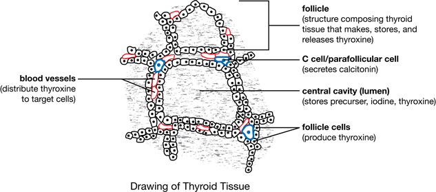
The structure of the thyroid gland is different from other endocrine glands. Thyroid tissue contains many spherical follicles consisting of a single layer of secretory cells surrounding a storage space. The secretory cells produce a precursor of thyroxine, which is stored in the storage space. Stimulation by thyroid stimulating hormone, which is produced by the anterior pituitary, causes the follicles to change the precursor and release it as thyroxine into the many blood capillaries between the follicles. Look at “Figure 13.16(C),” a micrograph of thyroid tissue, on page 448 of your textbook. Notice that there are some large cells (C cells or parafollicular cells) between the follicles. These cells produce and secrete calcitonin, another important thyroid hormone that will be addressed later in this lesson.
follicle: as it relates to the thyroid gland, a microscopic structure consisting of a circle of cells called follicle cells surrounding a central cavity
The thyroid gland consists of many follicles.
precursor: an inactive form of a molecule that can easily be changed into the active form
The precursor thyroglobin can easily be changed into the active thyroxine by the addition of iodine atoms.
thyroxine: a hormone produced by the follicle cells of the thyroid gland; mainly controls the rate at which the body metabolizes carbohydrates, fats, and proteins for energy and stimulates the proper development of the nervous system
C cells (parafollicular cells): cells located between the follicles of the thyroid gland that are responsible for synthesizing and secreting calcitonin
calcitonin: a hormone produced by the C cells (parafollicular cells) in the thyroid gland that decreases blood calcium levels by increasing uptake of calcium into the skeleton and inhibiting decomposition of bone when the blood levels of calcium are too high
Read “The Thyroid Gland: A Metabolic Thermostat” on pages 446 and 447 in your textbook. Study “Figure 13.16” on page 448, and then complete the following Self-Check activity.
 Self-Check
SC 1. On the diagram of thyroid tissues, label the follicle, storage space, C cells, and blood capillaries (vessels). Annotate the diagram by indicating the function of each structure, in brackets, after its label. Once you have finished, check your diagram and annotations and store it in your course folder.
Self-Check Answers
SC 1.