Module 2
1. Module 2
1.34. Page 2
Module 2—The Endocrine System
 Explore
The Anatomy of the Pancreas
pancreas: a gland with dual functions
The non-endocrine part secretes digestive enzymes into the intestine through the pancreatic duct, while the endocrine portion, called the islets of Langerhans, secretes insulin and glucagon into the bloodstream.
islets of Langerhans (islets): clusters of alpha and beta cells that secrete glucagon and insulin, respectively, into the blood
beta cells: cells in the islets that secrete insulin
insulin: a hormone secreted by the beta cells in the islets of the pancreas that lowers blood glucose levels by promoting the uptake of glucose by most cells of the body, and the synthesis and storage of glycogen in the liver; also stimulates protein and fat synthesis
alpha cells: cells in the islets that secrete glucagon
glucagon: a hormone secreted by the alpha cells in the islets of the pancreas that raises blood glucose levels by stimulating liver cells to convert glycogen to glucose; also stimulates fat cells (adipose cells) to convert fat to glucose
The pancreas is a dual-function organ located in the space between the stomach and the first loop of the small intestine. In Biology 20 you learned about the digestive enzymes it produces. Scattered among the enzyme-producing cells are clusters of cells called the islets of Langerhans (or simply islets), which produce pancreatic hormones. The vast majority of cells in the islets are beta cells, which produce insulin. A few elongated alpha cells produce glucagon. Examine the magnified section of “Figure13.24(B)” on page 456 of your textbook to locate these types of cells. (Note: The labels for the alpha and beta cells should be reversed in “Figure 13.24(B).” The beta cells are more numerous.)
Depending on your learning style, you may choose to complete the Read activity or the Watch and Listen activity that follows it.
 Read
Read
To give you a better understanding of the structures of the pancreas, read from the start of page 456 up to the heading “The Effects of Glucose Imbalance” on page 457 in your textbook.
 Watch and Listen
View the segment called “The Pancreas” from “The Pancreas: Regulating Blood Glucose Levels.” You may be required to enter a username and password to access the video. Contact your teacher for this information.
 Self-Check
You may choose to complete SC 1 or SC 2.
SC 1. To apply your knowledge of the anatomy of the pancreas and the function of its various structures, label the structures on the diagram of pancreatic tissue and associated structures. After you have labelled each structure, annotate the structures with their functions. Several structures are shown that you have studied in previous lessons. After you have completed the exercise, check your answers and file your work in your course folder.
Self-Check Answers
SC 1.
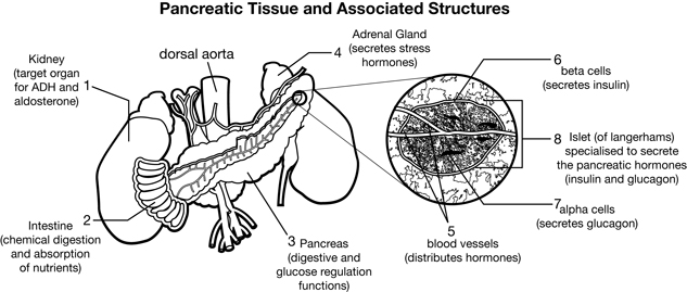
Inquiry into Biology (Whitby, ON: McGraw-Hill Ryerson, 2007), BLM 13.4.1. Reproduced by permission.
SC 2. Practise identifying and labelling the structures of the pancreas by completing this drag-and-drop exercise.