Module 3
1. Module 3
1.13. Page 3
Module 3—The Male and Female Reproductive Systems
 Module 3: Lesson 3 Assignment
Retrieve the copy of the Module 3: Lesson 3 Assignment that you saved to your computer earlier in this lesson.
Complete the three drawings and three questions in the Lesson 3 Assignment when instructed to do so as you complete the following lab. When complete, save your assignment in your course folder. You will receive instructions later in this lesson about when to submit your assignment to your teacher.
 Lab—Examining Gonads and Gametes
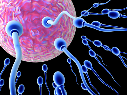
© Spectral-Design/shutterstock
Although the testes and ovaries are quite different structures, they serve the same two basic functions:
-
development of gametes
-
secretion of sex hormones
In this investigation you will compare ovarian and testicular tissue using microscopy (the scientific term for viewing objects with a microscope). From the images provided, you will identify the supporting structures that help develop the egg and sperm cells.
This lab is similar to the one on page 483 of the textbook. However, because you may not have a microscope, the microscope images will be provided to you. The procedure has been rewritten to guide you through the lab using the microscopic images.
Problem (Purpose)
How do the structures of testicular and ovarian tissues relate to their biological functions?
Materials
-
blank paper
-
pencil
-
microscopy images of testis: 100X, 200X, 400X (provided below)
-
microscopy images of cat follicle: 100X, 200X, 400X (provided below)
Procedure
Part 1—Testicular Tissue
Step 1: View the microscopy and other model images of the testes below. Where it is provided, pay attention to the magnification power given.
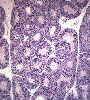
Microscope image of a cross-section of a seminiferous tubule at 100X magnification
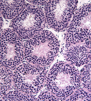
Microscope image of a cross-section of a seminiferous tubule at 200X magnification

Microscope image of a cross-section of a seminiferous tubule at 400X magnification
Step 2: Read “Appendix D: Review of Biological Drawings” on page 759 of the textbook. Follow the instructions given when making the biological drawings requested in this lab.
Step 3: Search the Internet for images of the internal structure of the testes. You could start your search using the phrase "seminiferous tubules male reproductive system anatomy." Use results from medical schools to study the internal structures.
Step 4: Examine the microscopy images provided above. Look for several circular structures. These are the seminiferous tubules. Try to identify the following cells in the images: lumen, seminiferous tubule wall, spermatogonial cells, spermatocytes, spermatids and spermatozoa or mature sperm cells, Sertoli cells, and interstitial cells.
- Draw a diagram of the specimen as it appears under the highest power magnification. Label the following cells in your drawing: lumen, seminiferous tubule wall, spermatogonial cells, spermatocytes, spermatids and spermatozoa, Sertoli cells, and interstitial cells. If possible, paste your image into the space provided for question 1 in your Lesson 3 Assignment document; otherwise, submit your drawing to your teacher when instructed to do so later in this lesson.
Part 2—Ovarian Tissue
Step 5: View the microscopy and other model images of the ovaries below. Where it is provided, pay attention to the magnification power given.
Step 6: Search the Internet for images of the internal structure of the ovary. You could start your search using the phrase "human female reproductive system histology." Use results from medical schools to study the internal structures.
Step 7: Examine the microscopy images provided below. Look for larger circular structures; these are developing egg cells. Try to identify the following structures in the images: primary follicle, primary oocyte, mature follicle, mature ovum, ovarian tissue, and corpus luteum.
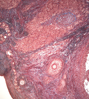
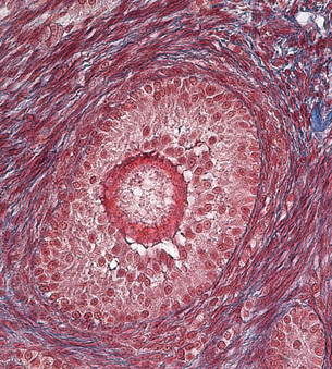
Microscope images of a cross-section of a cat ovary showing a primary follicle at 100X magnification
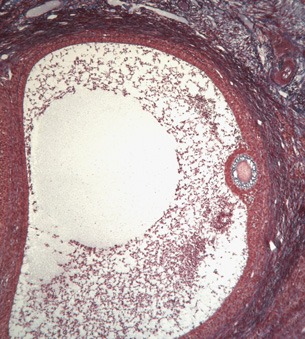
Microscope image of a cross-section of a cat ovary showing a view of a mature follicle at a 100X magnification
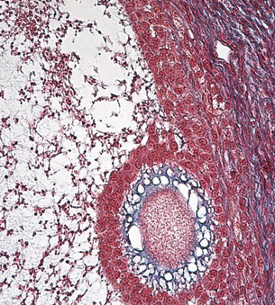
Microscope image of a cross-section of a cat ovary showing a view of a mature follicle at a 400X magnification
-
Draw a diagram of the specimen image, Cat, Primary Follicle, 100X. Label the following structures in your drawing: primary follicle, primary oocyte, ovarian tissue, and corpus luteum. If possible, paste your image into the space provided for question 2 in your Lesson 3 Assignment document. Otherwise, submit your drawing to your teacher when instructed to do so later in this lesson.
-
Draw a diagram of the specimen image, Cat, Mature Follicle, 100X. Label the mature oocytes and follicle cell structures in your drawing. If possible, paste your image into the space provided for question 3 in your Lesson 3 Assignment document. Otherwise, submit your drawing to your teacher when instructed to do so later in this lesson.
Observations
After examining the microscopy slides of testicular and ovarian tissues, you should have completed three diagrams with appropriate labels and titles. You should now be able to identify the functional gametes as well as their supporting structures.
Questions
Answer the questions 4 through 6 in the Lesson 3 Assignment document using knowledge from examining the gonad tissue sample images.
Conclusions
In this lab you identified the location and function of the gonads, gametes, and their supporting structures. Reproduction and, therefore, continuation of the species would be impossible if not for proper functioning of the supporting cells that develop the gametes.
Real-World Applications (Going Beyond)
In this lab you have seen gonad tissue and identified some of the structures within these organs. Things are never quite as simple as they seem; there are other structures that have not been mentioned in this lab or in your textbook. See if you can research and add the following structures to your diagrams: testicular basement membrane, blood-testis barrier, blood vessels, early spermatid, late spermatid, corpus albicans, early and mature corpus luteum, primordial follicles, follicular cavity, follicular fluid, zona pellucid, and corona radiate.
 Self-Check
Complete “Section 14.1 Review” questions 1 to 4 and 6 to 8 on page 485 of the textbook to see how well you understand the development of the egg and sperm cells.
Self-Check Answers
“Section 14.1 Review” Answers
- The two main purposes of the gonads are to produce gametes and to secrete sex hormones.
- a. fimbriae: found in the female reproductive system; it is responsible for helping to move the ovum (released during ovulation) into the oviduct. It sweeps over the ovary and moves the ovum into the cilia-lined oviduct.
b. ductus deferens: found in the male reproductive system; it is a storage duct responsible for storing and eventual transport of sperm to the urethra during ejaculation.
c. endometrium: found in the female reproductive system; it is the uterine lining that will support an implanted embryo.
d. epididymis: found in the male reproductive system, it stores the sperm during maturation and as they become motile. Once matured, the sperm move to the ductus deferens
- Sperm move out of the epididymis into the ductus deferens, where they are mixed with various fluids to make semen. The seminal vesicles provide a mucus-like fluid containing fructose for energy; the prostate gland and Cowper's gland provide alkaline and mucus-like fluids that can neutralize the acids in the female reproductive tract. The combination of sperm and the fluids make up semen.
- The ovum is moved from the ovary into the oviduct with the aid of the fimbriae, which are thread-like projections. As the egg moves down the oviduct it is aided by the beating of the cilia. This creates a current that moves the ovum towards the uterus.
- a. an oocyte
b. follicle
c. ovarian tissue
- The sperm is composed of three parts: a head, a mid-section, and a tail. The tail provides the motility required to move the sperm through the female reproductive tract; the tail is powered by a middle section that has mitochondria that can use the fructose provided by the seminal vesicles to make energy. The head section carries both the chromosomal material and the acrosome (a cap-like structure that contains the enzymes needed to penetrate the jelly-like layer surrounding the egg). The much larger, round ovum is covered by a specialized layer that only allows sperm with acrosome enzymes to penetrate. The egg has no structures to support motility, making it a more accessible target for the sperm, and its larger size makes it easier for the tiny sperm to attempt to fertilize it. The egg also contains cytoplasm and organelles to support the zygote as it makes its way into the uterus to implant itself in the endometrium.
- By wearing looser pants, his scrotum will be further away from his body and at a temperature more conducive to the production of viable sperm—35°C as opposed to the usual 37°C. High temperatures can cause deformed sperm to mature, as well as decrease the number of sperm produced.