Module 5
1. Module 5
1.13. Page 2
Module 5—Cell Division: The Processes of Mitosis and Meiosis
 Explore
 Read
Read
In previous lessons you learned how cells follow a cycle. From origin, through G1, S, and G2 of interphase, the cell grows, divides its DNA, and prepares for cell division.
Mitosis is an orderly process that carefully divides a cell’s chromosomes. These chromosomes are copied precisely in S phase so that each daughter cell receives the identical genetic content. When mitosis is complete, cytokinesis divides the cell physically into two identical daughter cells, which are exact replicas of the parent cell.
Mitosis is a process, and is actually more of a continuum than a set of snapshots. However, for study purposes, mitosis is divided into four distinct phases: prophase, metaphase, anaphase, and telophase. These phases are very important. PMAT is an easy mnemonic that can be used to remember the order of the phases and is a great study and memorization tool!
prophase: the first phase of mitosis where visible chromosomes appear scattered through a cell; nuclear membrane dissolves; centrioles move to opposite poles, forming a spindle between them
metaphase: the second phase of mitosis where chromosomes line up on the equator (metaphase plate) and attach via their centromeres to a spindle fibre
Each centromere replicates so each sister chromatid has its own to allow spindle fibre to attach.
anaphase: the third phase of mitosis where spindle fibres contract, pulling sister chromatids of each chromosome apart to opposite poles
telophase: the fourth phase of mitosis where nuclear membranes form around the two groups of chromosomes; spindle apparatus dissolves; chromosomes decondense to become chromatin
centrioles: organizing bodies of the spindle
As they move apart in prophase, spindle fibres stretch out between them, forming the spindle apparatus.
Read pages 557 and 558 and consider the summary of the phases in “Figure 16.8” on page 557 of your textbook. Summarize the information about these phases for your course folder. Study both the computer graphic and the actual slides of each phase that follow, noting the centrioles and spindle apparatus in prophase.
 Watch and Listen
You should now have functional knowledge of each of the phases of mitosis. Review how they work together by watching the animation “Mitosis and Cytokinesis.” Pay attention to key events and structures of each phase that will help you develop answers to the following questions:
- What happens to the nuclear membrane before, during, and after mitosis?
- What roles or actions do the spindle fibres fulfill?
- How do the chromosomes line up along the equatorial plate? Note: This animation uses the term kinetochore when referring to the centromere. Either is acceptable terminology.
Read
Consider the two slide images below. How are they similar? How are they different?

If you have good observation skills, you should notice two clear differences between plant and animal mitosis.
First, plant cell walls are rigid and cannot go through cleavage. Instead, a new cell wall is formed between the daughter cells. This is called a cell plate.
Second, plant cells do not have centrioles. They do form a spindle apparatus to move chromosomes around, but must anchor this apparatus to the cell wall instead of to the centrioles as animal cells do.
Read “Cytokinesis” and “Mitosis and Cytokinesis in Plant Cells” on pages 558 and 559 of your textbook. A table is an excellent tool for summarizing the differences between plant cell and animal cell cytokinesis.
 Module 5: Lesson 3 Assignment
Retrieve the copy of the Module 5: Lesson 3 Assignment that you saved to your computer earlier in this lesson. Complete the lab that follows, and answer the questions in the Lesson 3 Assignment. Save your work in your course folder. You will receive instructions later in this lesson about how to submit the assignment.
 Lab—Cell Division
You have two options for completing this lab: a lab simulation or an investigation from your textbook.
 Try This
TR 1. Follow the links below to two interactive diagrams of mitosis. Label the diagrams correctly.
 Self-Check
To apply your understanding, complete the following questions on mitosis and cellular division, and then check your answers. If you have questions or need clarification, consult with your teacher.
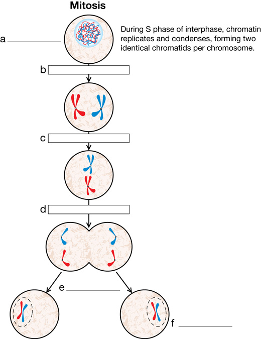
SC 1. Label the following terms on the flow chart below:
- mother cell in S phase of interphase
- daughter cells following cytokinesis
- anaphase
- metaphase
- prophase
- telophase
SC 2. A skin cell taken from a chimpanzee contains 48 chromosomes.
- How many chromosomes would there be in the nerve or bone cells of this animal?
- If a skin cell of the chimpanzee underwent cell division, how many chromosomes would there be in each daughter cell?
SC 3. What role do centrioles play in cell division of animal cells?
Stages |
||||
A. interphase |
B. prophase |
C. metaphase |
D. anaphase |
E. telephase |
Statements |
||||
________ a. |
Normal growth and functioning of the cell occurs here. |
|||
________ b. |
Chromosomes replicate to produce two sets of chromosomes in preparation for cell division. |
|||
________ c. |
Chromosomes with their duplicates still attached shorten by coiling, thus becoming visible under the microscope. |
|||
________ d. |
Centrioles migrate to opposite sides of the cell and the nuclear membrane dissolves. |
|||
________ e. |
Spindle fibres grow from each centriole and attach to the centromere of each chromatid pair. |
|||
________ f. |
Chromatid pairs still joined at the centromere line up along the middle of the cell, called the metaphase plate. |
|||
________ g. |
Chromatids are pulled apart by shortening of the spindle fibres. One complete set of chromosomes is pulled to each pole. |
|||
________ h. |
Chromosomes uncoil, spindle fibres dissolve, and cytoplasm divides. Two daughter cells are formed. |
|||
SC 5. Name the process of cytoplasmic division, and describe how it is different in plant and animal cells.
Self-Check Answers
SC 1.
- mother cell in S phase of interphase
- prophase
- metaphase
- anaphase
- telophase
- daughter cells following cytokinesis
SC 2.
- There would be 48 chromosomes in nerve and bone cells.
- There would be 48 chromosomes in each daughter cell.
SC 3.Centrioles provide attachment for spindle fibres and form the points to which chromatids are pulled during anaphase.
- A
- A
- B
- B
- B
- C
- D
- E
SC 5. Division of the cytoplasm is called cytokinesis. In animal cells, the cytoplasm pinches off, separating the two daughter cells. In plant cells, a new cell wall must form between the two nuclei because the existing cell walls are rigid and do not allow for pinching. The new cell wall is called a cell plate.