Module 5
1. Module 5
1.6. Page 4
Module 5—Wave Theory of Light
 Reflect and Connect
© Micah May/shutterstock
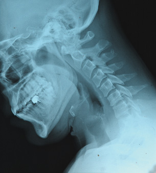
X-ray technologies are one of many diagnostics tools deployed in the health-care system. They also illustrate the production and transmission of electromagnetic waves.
An X-ray machine similar to those found in hospitals and dental offices uses a high voltage to accelerate an electron towards a tungsten metal target. With sufficiently high voltage, the electron gains enough energy to knock an inner shell electron out of the atom when it collides with the metal target. In an instant, an outer shell electron accelerates and replaces the lost inner electron to create an electromagnetic wave with the frequency and energy of an X-ray.
The wave then propagates outward toward the patient or specimen. Those waves that contact dense material—such as metal fillings, bones, and teeth—are absorbed and cast a shadow. Those waves that encounter less dense, soft tissues pass through and cause the white X-ray film to turn black and produce a negative image similar to the one in the photo.
Since X-rays have sufficient energy to knock out free electrons, they are considered a type of “ionizing radiation,” capable of damaging biological systems. Therefore, exposure to X-rays is minimized by taking images only when medically necessary and by protecting technicians by blocking X-rays with dense materials such as lead.
In the past, people did not always take such care around X-rays. In the 1940s and early 1950s, a shoe-fitting X-ray machine was a common fixture in shoe stores and department stores. These machines had three viewing ports so that your salesperson and a friend could look at your feet with you! The dangers of these machines became known in the 1950s; by the 1970s, their use was banned.
 Module 5: Lesson 1 Assignment
Remember to submit the Module 5: Lesson 1 Assignment to your teacher.