Module 1
| Site: | MoodleHUB.ca 🍁 |
| Course: | Biology 30 SS |
| Book: | Module 1 |
| Printed by: | Guest user |
| Date: | Friday, 19 December 2025, 4:20 AM |
Description
Created by IMSreader
Table of contents
- 1. Module 1
- 1.1. Big Picture
- 1.2. In this Module
- 1.3. Lesson 1
- 1.4. Page 2
- 1.5. Page 3
- 1.6. Page 4
- 1.7. Page 5
- 1.8. Lesson 2
- 1.9. Page 2
- 1.10. Page 3
- 1.11. Page 4
- 1.12. Page 5
- 1.13. Lesson 3
- 1.14. Page 2
- 1.15. Page 3
- 1.16. Page 4
- 1.17. Page 5
- 1.18. Page 6
- 1.19. Lesson 4
- 1.20. Page 2
- 1.21. Page 3
- 1.22. Page 4
- 1.23. Page 5
- 1.24. Page 6
- 1.25. Page 7
- 1.26. Lesson 5
- 1.27. Page 2
- 1.28. Page 3
- 1.29. Page 4
- 1.30. Page 5
- 1.31. Page 6
- 1.32. Page 7
- 1.33. Page 8
- 1.34. Lesson 6
- 1.35. Page 2
- 1.36. Page 3
- 1.37. Page 4
- 1.38. Page 5
- 1.39. Page 6
- 1.40. Page 7
- 1.41. Page 8
- 1.42. Page 9
- 1.43. Page 10
- 1.44. Page 11
- 1.45. Lesson 7
- 1.46. Page 2
- 1.47. Page 3
- 1.48. Page 4
- 1.49. Page 5
- 1.50. Page 6
- 1.51. Page 7
- 1.52. Lesson 8
- 1.53. Page 2
- 1.54. Page 3
- 1.55. Page 4
- 1.56. Page 5
- 1.57. Page 6
- 1.58. Page 7
- 1.59. Page 8
- 1.60. Page 9
- 1.61. Module Summary/Assessment
- 1.62. Module Glossary
1. Module 1
Module 1—The Nervous System
Introduction
In this module you will explore both the conscious and unconscious communication of your body. You will do this through an examination of the structure, organization, and function of the nervous system. You will discover how the nervous system works to maintain homeostasis, and you will learn what occurs when communications are disrupted or interrupted. You will research how imbalances and disorders cause the nervous system to function improperly and how medical technologies can be applied to correct these situations.
As you progress through the course, you will be encouraged to use science and technology to acquire new knowledge and to solve problems. The scope and characteristics of science, its connections to technology, and the social context in which it is developed will be examined. You will be asked to critically address science-related societal, economic, ethical, and environmental issues. The information in your textbook and the assessments in this module provide you with an opportunity to consider these issues related to the nervous system.
In the Biology 30 Course Introduction, several reference resources, such as The Key and Student Notes and Problems Workbook: Biology 30, were recommended to you for additional support. The multiple-choice, numerical-response, and written-response questions from past Diploma Exams are useful review and study resources. You will find these resources invaluable as you work through this module and the rest of Biology 30.
1.1. Big Picture
Module 1—The Nervous System
 Big Picture

© Monkey Business Images/shutterstock
You are at a gathering with your friends when, from across the room, you notice a girl who is new to your school. You can see her smile and can hear her laughter from across the room. You decide that you want to forget about your shyness and welcome her to your school, so you walk across the room to say “hi.” You notice that your heart is pounding, you’re breathing fast, and your hands are clammy. You are introduced and you shake hands. Without thinking, you yank your hand away from the handshake that’s hurting your sprained finger. You’re so embarrassed! Your body is communicating even if you don’t want it to! You stammer out a “hello” and she says “hello” back. You can see from her body language that she is nervous as well. Whether you end up with a new friend will ultimately be up to how you communicate, both intentionally and unintentionally.
While you may have felt like your nervous system was not working correctly because you could not control what your body was communicating, it was, in fact, responding and working to regain homeostasis, the balance necessary for the health and efficient function of your systems. The unconscious division of your peripheral nervous system brought your heart rate back to normal. You took a deep breath and, to some extent, controlled some of your body’s homeostatic responses.
In Module 1 you will learn about the many ways your nervous system communicates with your body to speed you up, slow you down, and establish homeostasis.
You may have heard of diseases, such as multiple sclerosis or Alzheimer’s disease, where the nervous system is not in homeostasis and system health has been compromised. In this module you will examine several diseases where communication in the nervous system has been interrupted.
In this module you will explore how the body communicates through the nervous system. To do this, you will need to explore the following focusing questions:
- How is the nervous system organized? How do its parts communicate with one another? What could interrupt this communication?
- What are the main structures and functions of the brain? How does the brain establish communication? What happens when this communication is interrupted?
- What are the main features of the spinal cord? What role does the spinal cord play in the communication and coordination of the rest of the body?
- What are the features of the building blocks of the nervous system?
- What information about the environment do the sensations of touch, smell, and taste communicate to the nervous system in order to maintain homeostasis?
- What are the major parts of the eye? How do they function? How do they support the integrated act of seeing?
- What are the major parts of the ear that facilitate your response to sound and your ability to maintain balance within the changing environment?
- How does the structure of a neuron facilitate the reception and transmission of a nerve impulse to the synaptic gap?
- What are the events in the synaptic gap that affect how neurons communicate with each other?
You have been introduced to the focusing questions for this module. Each lesson will restate these focusing questions to guide your study. To help you organize the concepts of this module, and to provide you with a potential aid for review, you may choose to download the Module 1 Concept Organizer. Fill in this concept organizer with the ideas that you master as you work through each lesson, or prepare the organizer when you have completed Module 1. You can use keywords, point form, or any amount of detail that meets your needs. You may choose to work from the file on your computer, print the document and work from the paper copy, or copy the outline onto a large sheet of poster paper. After you have prepared your mind map, you may wish to check your work with the concept organizer provided in the Module Summary. The concept organizer provided outlines some of the key topics that you should include in each lesson of your concept organizer. This is a great tool to review and use for study purposes, but using this organizer is completely your choice.
In the Module Assessment for Module 1, you will complete a detailed study of Alzheimer’s disease. In each lesson of Module 1, you will consider how that lesson’s topic could be related to the cause or symptoms of Alzheimer’s disease. You will complete some work towards the Module Assessment project in each lesson. For more details about the Module Assessment and the evaluation criteria, visit the Module Summary and Assessment section.
1.2. In this Module
Module 1—The Nervous System
In This Module
Inquiry Question: How does the nervous system communicate with the body and maintain homeostasis?
There are eight lessons in Module 1.
Most of the lessons are designed to take you 80 minutes to complete; however, some lessons may take longer because of the significance of the concept being covered in the lesson. The suggested lesson times do not include the time needed to complete such activities as “Try This,” “Watch and Listen,” assignments, practice questions, review, or research.
This module corresponds to Chapters 11 and 12, pages 360 to 433, in your textbook. Lessons 1, 2, 3, 7, and 8 correspond to Chapter 11; and Lessons 4 to 6 correspond to Chapter 12. Before you begin your study of Lessons 1 to 3, you may wish to read Chapter 11 for an overview. As you begin each lesson, you may wish to read the relevant chapter in the textbook for an overview before you begin.
Lesson 1: Structure and Organization of the Nervous System
Studying the structure and organization of the nervous system helps you understand how some responses can be controlled, whereas others just seem to happen.
In this lesson you will investigate the following focusing questions:
- How is the nervous system organized, and how do its parts communicate with each other?
- What interrupts the normal communication mechanisms of the sympathetic and parasympathetic parts of the nervous system?
Lesson 2: The Brain and Spinal Cord—the Boss and Unthinking Boss
The brain is the control centre for your body, just like the nucleus controls cell function. If the spinal cord is damaged, your brain can’t communicate successfully with your body.
In this lesson you will investigate the following focusing questions:
- What are the main structures of the brain and spinal cord, what are their functions, and how do they co-ordinate those functions?
- What happens when the information to or from the brain or spinal cord is disrupted or interrupted?
Lesson 3: The Neuron and the Reflex Arc
The neuron is the basic building block of the nervous system. Examination of the reflex arc illustrates the types of neurons and their role in communication.
This lesson helps you to understand the following focusing questions:
- What are the structures and functions of the neuron, and how do they support communication?
- What are the components of a reflex arc?
Lesson 4: Sensory Perception—Taste, Smell, Touch, and Temperature Sensations
Sensory perception helps you establish homeostasis but involves senses that are not considered to be as significant as vision and hearing. In this lesson you will examine the senses of taste, smell, touch, and temperature.
The following focusing question will be addressed:
- What information about the environment do the sensations of touch, smell, and taste communicate to the nervous system in order to maintain homeostasis?
Lesson 5: Photoreception—the Eye
Vision is the dominant sense. The structures of the eye facilitate vision (photoreception).
The following focusing question will help you to understand the concept of photoreception:
- What are the major parts of the eye, how do they function, and how do they communicate with the nervous system to support the integrated act of seeing?
Lesson 6: Mechanoreception—the Ear
It is said that the second most important sense is mechanoreception. The ear is the organ that accommodates both hearing and balance.
Understanding the following focusing questions will help you to learn about mechanoreception.
- What are the structures of the ear, and what are their functions in communicating sound?
- How does the ear impact your ability to maintain balance within your changing environment?
Lesson 7: The Nerve Impulse—Transporting the Message
In this lesson you will explore the path of a nerve impulse as it is communicated through the structures of the neuron. You will also examine what happens when communication is interrupted by disorders like multiple sclerosis.
You will investigate the following focusing question in this lesson.
- How does the structure of a neuron facilitate the reception and transmission of a nerve impulse to the synaptic gap?
Lesson 8: Synaptic and Neuromuscular Transmission—Crossing the Divide
Neurons are physically separated from each other by tiny gaps. The nerve impulse must be transmitted across these gaps by chemicals called neurotransmitters. Substances can alter events at the synaptic gap. Coffee can excite, whereas alcohol can inhibit, synaptic gap transmission.
To understand the concept of synaptic transmission, you will investigate the following focusing questions:
- How do the anatomy and function of the synaptic gap and neuromuscular junction facilitate the transmission of nerve impulses between neurons and between neurons and effectors?
- How do chemicals that are taken into your body and disorders, such as Parkinson’s disease, compromise synaptic transmission?
1.3. Lesson 1
Module 1—The Nervous System
Lesson 1—Structure and Organization of the Nervous System
 Get Focused
In the Big Picture you imagined being in a room full of people and trying to get the nerve to approach a student who is new to your school, and hopefully make a new friend. You started to breathe faster and your heart rate went up as you approached her to say “hi.” It took some effort to calm yourself down. In this situation, you could not control your breathing and heart rate, but you did have control of your legs and the direction in which they were taking you.
nervous system: an elaborate communication system that receives input; processes, integrates, and stores information; and triggers muscle contraction or glandular secretion
homeostasis: the tendency of the body to maintain a state of equilibrium or a stable internal environment
Can you imagine being able to control your breathing and heart rate like you control your legs? This is where the nervous system gets divided into the unconscious, or involuntary, section and the conscious, or voluntary, part. Processes vital to life, like breathing, are controlled unconsciously. Just try to see how long you can hold your breath! In this lesson you will explore mechanisms that increase your breathing rate and slow it down, returning you to a normal or balanced state. This state is called homeostasis.
Controlling your skeletal muscles, such as your legs, is a conscious or voluntary act. In Lesson 1 you will discover the part of the nervous system responsible for voluntary control, and you will see what happens when the pathways in your nervous system are interrupted.
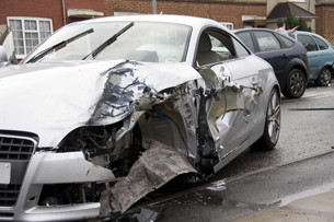
© Solid Web Designs LTD/shutterstock
Injuries, such as paralysis or the loss of a limb, can interrupt communication in the conscious part of your nervous system. However, someone seriously injured in a car accident may learn how to walk again through the re-establishment of communication in the conscious nervous system. This may be achieved through a combination of physiotherapy, surgery, and other technologies developed to enhance or repair communication pathways in the nervous system.
In this lesson you will investigate the following focusing questions:
- How is the nervous system organized, and how do its parts communicate with each other?
- What interrupts the normal communication mechanisms of the sympathetic and parasympathetic parts of the nervous system?
sympathetic nervous system: the division of the autonomic nervous system that activates the body to cope with some stressor, such as danger, excitement, or fear; sometimes referred to as the fight, fright, and flight subdivision
parasympathetic nervous system: the division of the autonomic nervous system that oversees digestion, elimination, and glandular function; often works opposite the sympathetic nervous system to bring the body back to normal
 Module 1: Lesson 1 Assignment
Your teacher-marked Module 1: Lesson 1 Assignment requires you to submit a response to the following:
-
Part 1—Response to Video
-
Part 2—Reflect and Connect
Download a copy of the Module 1: Lesson 1 Assignment to your computer now. You will receive further instructions about how to complete this assignment later in the lesson.
The other questions in this lesson are not marked by the teacher; however, you should still answer these questions. The Self-Check and Try This questions are placed in this lesson to help you review important information and build key concepts that may be applied in future lessons.
After a discussion with your teacher, you must decide what to do with the questions that are not part of your assignment. For example, you may decide to submit to your teacher the responses to Try This questions that are not marked. You should record the answers to all the questions in this lesson and place those answers in your course folder.
During this lesson you will begin the Module Assessment project that you were introduced to in the Big Picture. This will begin with initial research into Alzheimer’s disease. This research will be stored in your course folder.
1.4. Page 2
Module 1—The Nervous System
 Explore
 Watch and Listen
central nervous system (CNS): the part of the nervous system that includes the brain and spinal cord
peripheral nervous system (PNS): the portion of the nervous system consisting of nerves and ganglia (collections of nerve cell bodies) that are outside the brain and spinal cord
neuron: the basic functional cell of the nervous system that is specialized to generate and transmit nerve impulses (messages)
nerve: a message pathway of the nervous system; made up of many neurons grouped into bundles and surrounded by protective tissue
There are 12 pairs of cranial nerves that insert into the brain and 31 pairs of spinal nerves that emanate from the spinal cord.
View the video “Electrochemical Control Systems in Humans: Regulating Physiological Processes.” You may be required to enter a username and password in order to access the video. Contact your teacher for this information.
As you watch the video, note the two divisions of the nervous system—the central nervous system, which is composed of the brain and the spinal cord, and the peripheral nervous system. Watch for the divisions of the peripheral system and how they control the body consciously and unconsciously. You may wish to prepare a flow chart or diagram to illustrate these relationships for later review.
The video also demonstrates communication and homeostasis. It explains the structures of the neuron, which is the basic building block of the nervous system, and outlines how neurons aid communication.
The organization of nerves to support communication is also detailed in this video. As you explore typical communication, keep in mind what could result when interruptions occur in the nervous system due to diseases like multiple sclerosis and Alzheimer’s disease.
You may choose to make rough, point-form notes as you watch the video. Such notes will be helpful in preparing your work in later lessons that deal specifically with these topics from the video. Your notes will also be useful in completing your assignment.
As you watch the video, pay specific attention to the following topics:
- the description and examples of homeostasis
- all the divisions of the nervous system and the examples of what each division does
- the neuron structure and its organization into nerves
- the biological basis of multiple sclerosis
 Module 1: Lesson 1 Assignment
Retrieve the copy of the Module 1: Lesson 1 Assignment that you saved to your computer earlier in this lesson. Complete Part 1—Response to Video. Save your assignment in your course folder. You will receive instructions about when to submit your assignment to your teacher later in this lesson.
 Read
Read
The “Electrochemical Control Systems in Humans: Regulating Physiological Processes” video that you watched above provides a good introduction to the various parts of the nervous system and how they work. You may wish to read pages 366 to 369 and pages 396 to 399 in your textbook to increase your understanding. Based on your reading and the notes you made as you watched the video, make summary notes or a graphical organizer of this material to increase your understanding of the nervous system. Save your notes or graphical organizer in your course folder.

autonomic nervous system (ANS): a division of the peripheral nervous system that conducts nerve impulses to cardiac and smooth muscles, as well as to glands; may also be called the involuntary motor system
somatic nervous system (SNS): a division of the peripheral nervous system that conducts nerve messages to the skeletal muscles; may sometimes be called the voluntary nervous system
In both the video and the reading, the divisions of the nervous system, including the autonomic nervous system and the somatic nervous system, were shown to be vital to maintaining homeostasis.
The following Self-Check is an opportunity to practise your understanding of the autonomic and somatic nervous systems and how they communicate to maintain balance.
You can revisit the video or the reading if you need to review these systems.
 Self-Check
SC 1. Complete the Comparing the Somatic and Autonomic Nervous System Self-Check.
1.5. Page 3
Module 1—The Nervous System
 Reflect and Connect
You now have the opportunity to reflect on the parts of the nervous system and how they communicate to maintain homeostasis.
 Module 1: Lesson 1 Assignment
Retrieve the copy of the Module 1: Lesson 1 Assignment that you saved to your computer earlier in this lesson. Complete Part 2—Reflect and Connect. Save your completed assignment in your course folder. You will receive instructions about when to submit your assignment to your teacher later in this lesson.
 Self-Check
SC 2. Define homeostasis. Illustrate your answer by using body temperature as an example.
SC 3. Explain why the nervous system is critical for maintaining homeostasis.
SC 4. Identify what basic neural pathway is involved as you dodge a misdirected tennis ball.
SC 5. How is the autonomic nervous system different from the somatic nervous system?
SC 6. Why do you have to learn how to walk but not how to breathe?
Self-Check Answers
SC 2. Homeostasis is the maintenance of a nearly constant internal environment that fluctuates about a set point, or an ideal point. For example, the set point for body temperature is 37°C. However, the temperature hovers around that value, either below or above by a small amount. When a deviation from the set point occurs, the nervous system will take corrective action; for example, shivering to generate heat in response to feeling cold.
SC 3. The human nervous system is capable of regulating thousands of activities simultaneously. The overall function of the nervous system is to collect information, analyze (sort, integrate, and interpret) the information, and to initiate an appropriate response to maintain a state of dynamic equilibrium or homeostasis. The nervous system accomplishes this function quickly and precisely. Quick action is often needed to get you out of dangerous situations.
SC 4. A sense organ, in this case the eye, detects the ball and the sensory neuron (optic nerve) carries the message to the brain. At this point, an interneuron interprets the information and sends a message via a motor neuron, which causes muscles to act to withdraw (to move the head out of the way of the ball).
SC 5. The somatic nervous system, which takes messages via motor neurons to the skeletal muscles of the body, is controlled by the conscious part of the brain (cerebrum) and causes voluntary (conscious) movements of the muscles. The autonomic nervous system, which is composed of sympathetic and parasympathetic motor neurons, carries messages to cardiac and smooth muscles. This causes an involuntary (not necessarily conscious) contraction of these muscles.
SC 6. A person needs to learn how to contract skeletal muscles (voluntary control by the somatic nervous system). However, one does not need to learn how to contract smooth or cardiac muscle (involuntary control by the autonomic nervous system) or how to carry out reflexes, which are automatic.
1.6. Page 4
Module 1—The Nervous System
 Reflect on the Big Picture
In the Big Picture you imagined being at a gathering where you experienced all sorts of nervous responses in your body. In Lesson 1 you explored the unconscious and conscious communication of your nervous system.
The Module Assessment for Module 1 will require you to research Alzheimer’s disease. For full details about this project and the assessment criteria, go to the Module Summary and Assessment section.
You will begin this project now by researching Alzheimer’s disease to identify the affected parts of the nervous system and what implications there are to standard communication.
- Conduct an Internet search using the search terms or keywords “Alzheimer’s disease,” “Alzheimer’s + brain or neuron,” and “Alzheimer’s research.”
- Visit a local doctor’s office or regional health centre for publications about Alzheimer’s disease.
- Visit your local library.
 Module 1: Lesson 1 Assignment
Submit your completed Module 1: Lesson 1 Assignment to your teacher for assessment.
1.7. Page 5
Module 1—The Nervous System
 Lesson Summary
In this lesson you investigated the following focusing questions:
-
How is the nervous system organized, and how do its parts communicate with each other?
-
What interrupts the normal communication mechanisms of the sympathetic and parasympathetic parts of the nervous system?
To answer these questions, you explored the human nervous system as a complex communication system organized into the central nervous system and the peripheral nervous system. These systems work together to maintain homeostasis.
The motor neurons of the peripheral nervous system take information from the brain and spinal cord to the somatic nervous system and the autonomic nervous system. The autonomic nervous system is composed of two subdivisions—the parasympathetic and sympathetic systems.
The functional unit of the nervous system is the neuron. Neurons are bundled together to form nerves. The nervous system gathers information using sensory neurons or a sensory pathway. The nervous system integrates information using interneurons like those found in the brain and spinal cord. Instructions are then transmitted by motor neurons or motor pathways to muscles and glands. These muscles and glands are called effectors because they initiate a response.
These pathways may sometimes be disrupted, causing communication to be interrupted. In Lesson 3 of this module, you will explore an example of a rapid form of communication called a reflex arc. In other lessons you will continue to explore the consequences when communication in these pathways is interrupted.
In multiple sclerosis, a special type of communication pathway is disrupted so that messages get lost or reach muscles very slowly or sporadically. In Alzheimer’s disease, brain neurons become dysfunctional and nerve messages cannot be transmitted within the brain.
Lesson Glossary
Consult the glossary in the textbook for other definitions that you may need to complete your work.
autonomic nervous system (ANS): a division of the peripheral nervous system that conducts nerve impulses to cardiac and smooth muscles, as well as to glands; may also be called the involuntary motor system
central nervous system (CNS): the part of the nervous system that includes the brain and spinal cord
homeostasis: a state of body equilibrium or a stable internal environment of the body
nerve: a message pathway of the nervous system; made up of many neurons grouped into bundles and surrounded by protective tissue
There are 12 pairs of cranial nerves that insert into the brain and 31 pairs of spinal nerves that emanate from the spinal cord.
nervous system: an elaborate communication system that receives input; processes, integrates, and stores information; and triggers muscle contraction or glandular secretion
neuron: the basic functional cell of the nervous system that is specialized to generate and transmit nerve impulses (messages)
parasympathetic nervous system: the division of the autonomic nervous system that oversees digestion, elimination, and glandular function; often works opposite the sympathetic nervous system to bring the body back to normal
peripheral nervous system (PNS): the portion of the nervous system consisting of nerves and ganglia (collections of nerve cell bodies) that are outside the brain and spinal cord
somatic nervous system (SNS): a division of the peripheral nervous system that conducts nerve messages to the skeletal muscles; may sometimes be called the voluntary nervous system
sympathetic nervous system: the division of the autonomic nervous system that activates the body to cope with some stressor, such as danger, excitement, or fear; sometimes referred to as the fight, fright, and flight subdivision
1.8. Lesson 2
Module 1—The Nervous System
Lesson 2—the Brain and Spinal Cord—the Boss and Unthinking Boss
 Get Focused

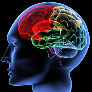
© Michael Monu/iStockphoto
Everyone has a brain and a spinal cord. Most people are born with both working the way they should. The brain is the centre of “who” you are—your personality, your values, your intellect, and your ability to determine right from wrong. Your brain is also the central controller of what you can do. It communicates with your spinal cord, allowing you to walk, move, feel, experience life, and perform body functions (like breathing).
Think back to the Big Picture. When you walked across the room to meet your potential new friend, it was your brain that projected your personality and your spinal cord that got you there.
In this lesson you will explore the relationships between the brain and the spinal cord, and you will see how dependant they are on each other to function correctly. When communication between the brain and spinal cord is interrupted, bad things happen. For example, your personality could change, you might lose the ability to walk, or your senses could be altered. Each of these is an example of interrupted communication.
In this lesson you will investigate the following focusing questions:
- What are the main structures of the brain and spinal cord, what are their functions, and how are these functions co-ordinated?
- What happens when the information to or from the brain or spinal cord is disrupted or interrupted?
 Module 1: Lesson 2 Assignment
Your teacher-marked Module 1: Lesson 2 Assignment requires you to submit a response to the following:
- LAB questions—LAB 1 or LAB 2
- Discussion questions—D1, D2, and D3
- Reflect and Connect—RC 1 or RC 2
Download a copy of the Module 1: Lesson 2 Assignment to your computer now. You will receive further instructions about how to complete this assignment later in the lesson.
You must decide what to do with the questions that are not marked by the teacher.
Remember that these questions provide you with the practice and feedback that you need to successfully complete this course. You should respond to all of the questions and place those answers in your course folder.
In Lesson 2 you will continue to work on the Module Assessment project by researching the relationship between Alzheimer’s disease, the brain, and the spinal cord. Your research will be stored in your course folder.
Required Materials and Equipment
- a sheep's brain
- rubber gloves
- safety goggles
- dissecting tray or foam tray
- sharp scissors with pointed tips
- sharp knife or scalpel
- a bleach solution to be used for cleanup
1.9. Page 2
Module 1—The Nervous System
 Explore
 Read
Read
© YAKOBCHUK VASYL /shutterstock
The brain is the control centre of your body. To understand where the brain processes the sight of a friendly face across the room or the sound of laughter, read from the heading “The Brain” on page 386 to the questions at the bottom of page 395 in your textbook. This will seem like a significant reading assignment, but concentrate your efforts on the following:
- the regions of the brain and their functions (The labelled diagram, “Figure 11.24,” on page 387 will further your understanding.)
- the lobes of the brain and the information they process
- technologies associated with understanding brain function, injuries, and diseases
 Watch and Listen
The following videos will provide an excellent introduction to the concepts that will be presented in this lesson. You may wish to return to these videos as you complete future lessons.
- “The Central Nervous System and Brain: Orchestrating Life”
- “The Peripheral Nervous System: Conscious vs. Unconscious Responses”
You may be required to enter a username and password to access these videos. Contact your teacher for this information.
 Self-Check
occipital lobe: one of the four lobes of the cerebrum that receives and analyzes visual information that is sent to association centres for recognition of what is being seen
SC 1. After reading pages 386 to 395 in your textbook and watching “The Central Nervous System and Brain: Orchestrating Life,” you will know that the occipital lobe processes sight and the temporal lobe processes sound. There are, however, four major lobes of the brain.
This diagram of the brain provides information about the functions of all four lobes. Consider this information as you identify and label the name for each lobe. After you have labelled the diagram, save it in your course folder for reference.
Self-Check Answers
SC 1. You should have labelled your diagram as follows.
- frontal lobe
- parietal lobe
- occipital lobe
- temporal lobe
SC 2. Check your understanding by answering questions 1, 4, 5, and 6 on page 395 of your textbook. The answers will help you build an understanding of key concepts.
You may choose to answer the easier questions mentally, and only record the answers to the questions that you find challenging. Or, you may decide to record your answers to all of the questions. The choices you make will depend upon the recommendation of your teacher, your learning style, your abilities in this subject, and your goals for the course. Verify your answers with your teacher.
 Module 1: Lesson 2 Assignment
Retrieve the copy of the Module 1: Lesson 2 Assignment that you saved to your computer earlier in this lesson. Complete Part 1—Labs. Save your assignment in your course folder. You will receive instructions about when to submit your assignment to your teacher later in this lesson.
1.10. Page 3
Module 1—The Nervous System
 Read
Read
© Stephen Sweet/shutterstock
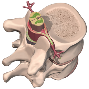
You must know the structure of the spinal cord to understand how it performs its role in the nervous system. Read from the start of page 385 to the heading “The Brain” on page 386 in your textbook. Make brief summary notes or draw a graphical organizer. Store your notes or graphical organizer in your course folder.
The spinal cord is the major communication link between the brain and the peripheral nervous system (PNS). As you learned by reading the textbook and watching “The Peripheral Nervous System: Conscious vs. Unconscious Responses,” the spinal cord is protected by the backbone and cerebrospinal fluid. You will learn more about the role of cerebrospinal fluid later in this lesson.
The spinal cord is made up of two special types of nerves.
- Sensory nerves communicate messages from the body to the brain for interpretation.
- Motor nerves communicate messages from the brain to effectors that initiate a response.
In Lesson 3 you will learn more about these communication pathways, and you will discover another communication role of the spinal cord—the reflex arc.
 Self-Check
SC 3. Complete this drag-and-drop activity to test your knowledge of the parts of the spinal cord.
The Meninges and Cerebrospinal Fluid
 Try This
TR 1. The brain and spinal cord are essential to communication and keeping your body in balance. They must be protected. This activity reviews the roles of the skull, meninges, and cerebrospinal fluid and illustrates what happens when the brain gets “scrambled.”
Find two plastic containers that are a little bit larger than an egg. Make sure these containers have lids. Put an egg into each container. Fill one container with water. Firmly close each container with its lid. Shake the containers. Make your observations. Consider what parts of this demonstration illustrate the skull, the meninges, the cerebrospinal fluid, and the brain. What was the role of the water?
TR 2. Complete questions 2, 3, and 7 on page 395 of your textbook. You may wish to discuss your answers with your teacher or post your answers for discussion. Save your work in your course folder.
Self-Check
These questions provide the opportunity to review and evaluate your understanding of the concepts in Lesson 2. Complete the questions, check your answers, and save your work in your course folder for reference when you study.
SC 4. The old adage “an elephant never forgets” appears to have some scientific basis. What area of the brain would you examine to begin researching this adage? Describe two technologies that might be useful in this research.
SC 5. Five situations in which you may find yourself are described below. Identify the part of the brain involved in processing each situation.
- You are on a boat. A sudden wind comes up, causing the boat to rock violently. You feel dizzy and nauseated, and you want to vomit.
- You are in a restaurant. The waiter brings you the bill. You reach into your pocket to retrieve your wallet.
- You are on a hike in the mountains when you suddenly come upon a grizzly sow with her two cubs. She rears up on her hind legs and growls menacingly. Your heart rate and breathing rate increase dramatically.
- You see a backpack.
- You hear a dog barking.
SC 6. Use the diagram of a neural pathway through the spinal cord to answer the questions that follow.
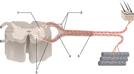
- Explain what would happen if only structure 2 were cut.
- Explain what would happen if only structure 4 were cut.
- Explain what would happen if structure 5 were cut.
Self-Check Answers
SC 4. The frontal lobe of the cerebrum appears to be where memory is created. In trying to investigate memory, MRI, PET, and CAT-scan technology may be used. Compare your description to the discussion on pages 392 to 394 of your textbook.
SC 5.
- cerebellum
- motor cortex of frontal lobe of cerebrum, or just cerebrum
- medulla oblongata and hypothalamus, or just medulla oblongata
- occipital lobe of cerebrum, or just occipital lobe
- temporal lobe of cerebrum, or just temporal lobe
SC 6.
- If only structure 2 (the dorsal root, which contains the sensory neurons) were cut, the person would not be able to sense stimuli but would be able to send nerve messages to the muscles.
- If only structure 4 (the ventral root, which contains the motor neurons) were cut, the person would not be able to send messages to the muscles—essentially paralyzing the muscles—but would be able to sense stimuli.
- If structure 5 (the spinal nerve, which contains both sensory and motor neurons) were cut, no sensory stimuli could be received from that area and no motor impulses could be sent to the muscles of that area.
 Discuss
Are you right-handed or left-handed? Which hand do you write with? For most people, the hand that they use for most activities is an indication of the dominant hemisphere of their brain. Research the activities that are controlled by the right and left hemispheres of the brain and how information is communicated between the hemispheres. Search terms that you could use include “right hemisphere,” “left hemisphere,” and “dominance in the brain.”
 Module 1: Lesson 2 Assignment
Retrieve the copy of the Module 1: Lesson 2 Assignment that you saved to your computer earlier in this lesson. In this part of your assignment, you will have the opportunity to discuss the role and characteristics of the dominant hemisphere of the brain. Complete Part 2—Discuss. Save your assignment in your course folder. You will receive instructions about when to submit your assignment to your teacher later in this lesson.
1.11. Page 4
Module 1—The Nervous System
 Reflect and Connect
You will now do some more work toward your Module Assessment. You will be able to choose between “shaken baby syndrome” and brain injuries related to not using a helmet. You will reflect on all that you have learned about the brain and spinal cord, and you will connect your understanding to the scenario that you choose to complete.
 Module 1: Lesson 2 Assignment
Retrieve the copy of the Module 1: Lesson 2 Assignment that you saved to your computer earlier in this lesson. Complete Part 3—Reflect and Connect. Save your completed assignment in your course folder. You will receive instructions about when to submit your assignment to your teacher later in this lesson.
Reflect on the Big Picture
In this lesson you examined how the sensations from your eyes and ears are communicated to the cerebrum of the brain and the occipital and temporal lobes for interpretation. You have examined the medulla oblongata and its role in communicating automatic, involuntary responses, such as the control of your breathing rate and heart rate. You have considered the role the spinal cord plays in communication, and you learned about the parts of the nervous system that protect these vital structures so that communication continues and homeostasis is maintained.
The Module Assessment for Module 1 involves a research project about Alzheimer’s disease. This disease is characterized by interruptions in communication within the nervous system. You have begun to collect information about this disease. Consider the information that you have in your course folder, and add any new information you learned in this lesson. Consider how Alzheimer’s disease affects
- the parts of the brain
- communication within the brain
- the function of the spinal cord
Store this additional research in your course folder to prepare for your Module Assessment.
Module 1: Lesson 2 Assignment
Submit your completed Module 1: Lesson 2 Assignment to your teacher for assessment.
1.12. Page 5
Module 1—The Nervous System
 Lesson Summary
In this lesson you have explored the following focusing questions:
- What are the main structures of the brain and spinal cord, what are their functions, and how are these functions co-ordinated?
- What happens when the information to or from the brain or spinal cord is disrupted or interrupted?
You have examined how all information about the external and internal environment is sent to the central nervous system, which consists of the brain and the spinal cord. Various part of the brain, including the cerebral hemispheres, the four lobes of the cerebrum, the cerebellum, the medulla oblongata, the pons, and hypothalamus, are responsible for receiving, sorting, interpreting, and co-ordinating information. The central nervous system (CNS) also initiates action in either the somatic or autonomic nervous system.
The spinal cord functions to receive sensory information via the sensory neurons in the dorsal root and either relays the messages to the brain or initiates an action through a motor neuron in the ventral root. In Lesson 3 you will learn about these neurons and the reflex arc, a major function of the spinal cord.
The destruction of the insulating layer around the neurons in the central nervous system (CNS) and peripheral nervous system (PNS) results in nerve impulses not being transmitted quickly enough or not being transmitted at all. This disorder is known as multiple sclerosis (MS). Muscle response becomes sporadic and eventually ceases. As you work through this module, you will understand the cause and symptoms of MS.
Lesson Glossary
Consult the glossary in the textbook for other definitions that you may need to complete your work.
occipital lobe: one of the four lobes of the cerebrum that receives and analyzes visual information that is sent to association centres for recognition of what is being seen
1.13. Lesson 3
Module 1—The Nervous System
Lesson 3—the Neuron and the Reflex Arc
 Get Focused
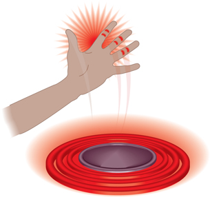
In the Big Picture you were introduced to the new girl in school, who you hoped would be your new friend. You shook hands with her when you were introduced, but your response to the handshake surprised you! You withdrew your hand right away—the finger you sprained in basketball practice hurt. How embarrassing!
reflex arc: an involuntary neural pathway that consists of a sensory receptor, a sensory neuron, a control centre that can be either the brain or the spinal cord, a motor neuron, and an effector that results in a reflex behaviour that usually has survival value
It may have been embarrassing, but your body knows what it’s doing. This immediate “no-brainer” response is exactly that—a “no brainer.” Your brain is not involved in this response. You didn’t think about the response. Instead, your spinal cord immediately responded to a “danger” signal and sent messages to your body for appropriate, fast responses to minimize tissue damage. This is called a reflex arc.
Other examples include pulling your hand away from a hot stove, quickly lifting your foot when you step on a tack, or swinging your leg up when the doctor taps your knee with a reflex hammer. In these cases, there is an immediate response—the “no-brainer” response. You jerk your hand away, lift your foot, or swing your leg. Then, other responses follow shortly afterwards. You may be embarrassed, cry out in pain, or respond in some other way. In these later responses, your brain is involved in processing information.
Why is the initial reaction so fast? Why did you become aware that the handshake hurt or that the stove was hot and respond afterwards?
neuron: a cell in the nervous system that generates and transmits nerve impulses; consists of dendrites, a cell body containing the nucleus, and an axon that may or may not have a myelin sheath
sensory neuron: a type of nerve cell that receives stimuli and conducts an impulse toward the brain and the spinal cord (central nervous system)
interneuron: a type of nerve cell found either in the brain or the spinal cord that transmits nerve impulses from sensory neurons within the various parts of the brain or to motor neurons
motor neuron: a type of nerve cell that transmits nerve impulses toward an effector, which can be a muscle or a gland
effector: one of the three types of muscle or a gland that responds to a nerve impulse
In this lesson you will study more about these types of reactions and the supporting structure and function of the neuron. Neurons are the specialized cells of the nervous system. These cells make it possible for you to see a person across the room, hear laughter, and smell a fragrance. You will examine how the neuron’s design can aid the speed of communication and how changes in its structure can result in an interruption in communication.
In Lesson 1 you learned about the three basic types of neurons and how each one carries out a specific function in the basic neural pathway. Sensory neurons communicate messages from the body to the central nervous system. Interneurons process information and communicate messages through motor neurons to effectors to produce a response. In this lesson you will examine the features of a neural pathway called the reflex arc. You will come to understand how this communication pathway produces extremely rapid responses that protect you and ensure your survival.
This lesson helps you to understand the following focusing questions:
- What are the structures and functions of the neuron? How do they support communication?
- What are the components of the reflex arc?
 Module 1: Lesson 3 Assignment
Your teacher-marked Module 1: Lesson 3 Assignment requires you to submit a response to the following:
- Lab Part 1
- Lab Part 2
Download a copy of the Module 1: Lesson 3 Assignment to your computer now. You will receive further instructions about how to complete this assignment later in the lesson.
You must decide what to do with the questions that are not marked by the teacher.
Remember that these questions provide you with the practice and feedback that you need to successfully complete this course. You should respond to all of the questions and place those answers in your course folder.
In Lesson 3 you will continue to work on your Module Assessment project by researching how the changes in the structure and function of the neuron could lead to the communication problems that exist in Alzheimer’s disease. You will consider whether or not there are any changes in the reflex arcs of patients with this disease. Your research will be stored in your course folder.
Required Materials and Equipment
In order to complete one of the labs, you will need cotton balls and a 20 cm x 20 cm clear plastic sheet.
1.14. Page 2
Module 1—The Nervous System
 Explore
In Lesson 1 you learned about the divisions of the nervous system that sent information to the brain for processing.
In Lesson 2 you learned the specific parts of the brain that are responsible for processing information. You also learned that the brain communicates with structures in your body using motor neurons to produce a response.
In Lesson 3 you will learn about the structures and functions of the neuron, the basic cell of the nervous system.
 Read
Read
To help you understand neurons, read “The Structure of a Neuron” on pages 370 and 372 in your textbook. You may wish to make summary notes and save them in your course folder for future study. It is important to include a labelled diagram of a neuron similar to “Figure 11.9” on page 372 of your textbook.
dendrite: a short, branching terminal of a neuron that receives input from other neurons or sensory receptors and transmits a nerve impulse toward the cell body
cell body: the part of a neuron that contains the nucleus and other cell organelles for carrying out the metabolic reactions of the nerve cell; relays the nerve impulse from the dendrites to the axon
axon: the long extension that emerges from the cell body and conducts the nerve impulse away from the cell body
The axon may be up to 1 m long in motor neurons.
Schwann cell: a type of supporting nerve cell that is found in the peripheral nervous system and wraps around axons of neurons and produces the myelin sheath
myelin sheath: a fatty insulating layer that surrounds axons and greatly increases the rate of impulse transmission and maintains the strength of the impulse by preventing the loss of ions along the length of the axon
node of Ranvier: a tiny gap in the myelin sheath surrounding the axon of myelinated neurons
Nerve impulse transmission occurs between nodes of Ranvier in what is called salutatory conduction, which causes the increase in the speed of impulse transmission.
axon terminal: numerous endings found at the end of an axon
Axon terminals are enlarged into knobs that are specialized for producing, storing, and releasing neurotransmitters.
You are now familiar with the dendrites, the parts of the nerve that receive impulses; the cell body, which carries out the life functions of the nerve cell; and the axon, which takes nerve impulses away from the cell.
Motor neurons communicate nerve impulses rapidly to effectors. Motor neurons are myelinated, which means their axons are surrounded by Schwann cells. Schwann cells produce a fatty material called the myelin sheath, which insulates the axon. This insulation prevents the loss of ions into the extracellular environment. You will learn about these ions and their role later in the module.
You will also learn how the nodes of Ranvier help to increase the transmission speed of nerve impulses and how the axon terminals produce neurotransmitters that complete communication between neurons. These neurotransmitters are necessary because neurons are not physically joined together. It is important to understand the basic structure and the function of the neuron in this lesson to help you master these more complex concepts.
In the nervous system, there are no physical connections between neurons to facilitate the transmission of an impulse. In electricity, the connections must be physically complete for the communication, or flow, of electricity to occur.

Adapted from Inquiry into Biology (Whitby, ON: McGraw-Hill Ryerson, 2007), 372, fig 11.9. Reproduced by permission
 Watch and Listen
To review the structures of the neuron, watch “Bio Reports: Neuron Structure and Function” in the video “Electrochemical Control Systems in Humans: Regulating Physiological Processes.”
 Self-Check
You should now be able to describe the three types of neurons and describe the structures and functions of the parts of a neuron.
SC 1. Complete the matching worksheet to show your understanding of the function and structure of neurons.
Self-Check Answers
SC 1.
Structure 1
Name: cell body
Function: performs life functions, relays messages to the axon
Structure 2
Name: nucleus
Function: control centre of the cell
Structure 3
Name: dendrites
Function: receive input from other neurons of sensory receptors and transmit toward the cell body
Structure 4
Name: myelin sheath
Function: fatty insulating layer that increases rate of communication transmission
Structure 5
Name: node of Ranvier
Function: gaps in myelin sheath that increase rate of communication transmission
Structure 6
Name: axon
Function: transmits nerve impulses away from cell body
Structure 7
Name: Schwann cellFunction: a type of supporting nerve cell that wraps around axons in the peripheral nervous system and produces myelin
 Try This
Check whether you can correctly assemble a neuron from a mad scientist’s tray of parts.
1.15. Page 3
Module 1—The Nervous System
 Module 1: Lesson 3 Assignment
In this part of your assignment, you will examine the structure of neural tissue and complete some questions related to function.
Retrieve the copy of the Module 1: Lesson 3 Assignment that you saved to your computer earlier in this lesson. Complete Lab Part 1. Save your completed assignment in your course folder. You will receive instructions about when to submit your assignment to your teacher later in this lesson.
 Try This
To enrich your understanding of the structures and their functions, try the Three Major Types of Neurons Drag-and-Drop. Drag each term to the correct area of the table.
Reflex Arcs
 Read
Read
To understand how the structures and functions of the neuron are related to the reflex arc, read “The Reflex Arc” on pages 369 and 370 in your textbook. As you read, create a labelled diagram of the neurons and the steps in a reflex arc.
In some sports, you need to be fast. In hockey, for example, the forward has to be faster than the defender in order to score. The goalie has to react quickly to stop the puck. Does the goalie think about how to stop the puck, or does the goalie just do it automatically?
reflex: an inborn, unlearned behaviour that results from the stimulation of a special neural pathway called the reflex arc
In the Module 1 Big Picture, you imagined being introduced to a new student in your school. Do you remember withdrawing your hand because the handshake hurt? After you thought about what happened, you were embarrassed. Some neurons are organized into special neural pathways that allow you to react before you are consciously aware of what is happening. These unlearned, unconscious, and very rapid pathways are called reflex arcs. The resulting behaviour is called a reflex. Do the following lab assignment to understand more about reflex arcs.
Module 1: Lesson 3 Assignment
In this part of the assignment, you will complete a lab studying the reflex arc.
Retrieve the copy of the Module 1: Lesson 3 Assignment that you saved to your computer earlier in this lesson. Complete Lab Part 2. Save your completed assignment in your course folder. You will receive instructions about when to submit your assignment to your teacher later in this lesson.
1.16. Page 4
Module 1—The Nervous System
 Watch and Listen
If you would like more review of reflexes, watch the following segments of the video “Reflexes and Synaptic Transmission: Getting the Message Across.”
- “Introduction”
- “Patellar and Pupillary Reflex”
- “Bio Probe: Checkstop”
- “Is Response Time Affected by Different Types of Stimuli?”
- “Bio Review: Conditioned vs. Simple Reflexes”
- “Reflex Arc”
- “Description of a Simple Reflex Arc”
- “Bio Fact: Typical Reflex Arc”
You may be prompted to provide a username and password in order to access the video. Contact your teacher for this information.
You can also check out an interesting applet about reflexes on the BBC website. You can use search words such as “bbc,” “schools,” “bite size,” and “bireflexarc.”
 Self-Check
SC 2. To review your understanding of the reflex arc, write a step-by-step description of the process illustrated below using all the terms that you have learned, and then name the type of reflex involved. Also provide a description of each numbered part of the diagram.
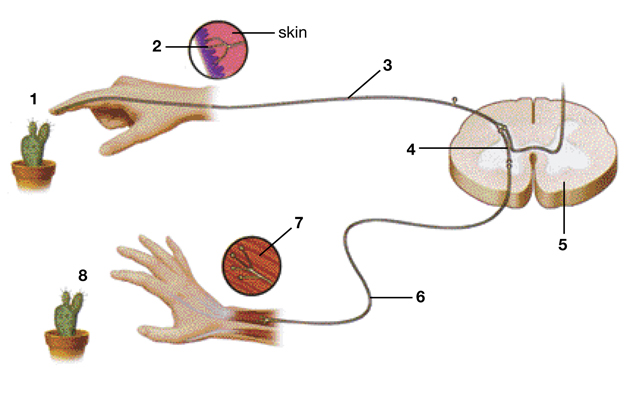
Inquiry into Biology (Whitby, ON: McGraw-Hill Ryerson, 2007), 370, fig. 11.8. Reproduced by permission.
Self-Check Answers
SC 2.
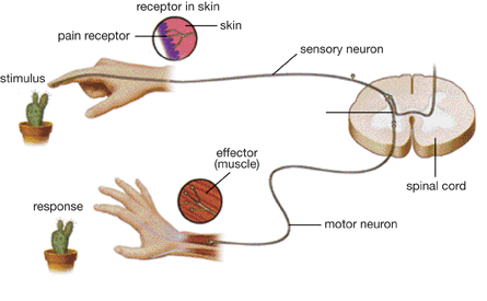
A withdrawal reflex is shown. Receptors in the skin perceive the stimulus (the cactus). Sensory information is conducted from the senses into the spinal cord. Motor information is then conducted away from the spinal cord to the muscles and glands.
1.17. Page 5
Module 1—The Nervous System
 Reflect and Connect
When you were introduced to the new girl at school, you quickly withdrew your hand from the handshake. You then became embarrassed and blushed. To reflect on your knowledge from this lesson, trace the reflex arc that occurred when you withdrew your hand. Include a description of all the parts of the motor neuron that communicated with your muscles. Also include a description of the role of the myelin sheath. Reflect on why your embarrassment and blushing happened later.
 Reflect on the Big Picture
In this lesson you learned about the structures and functions of the neurons. You also learned about the reflex arc. Research the possible connection between the cause and symptoms of Alzheimer’s disease and the parts of a neuron. Alzheimer’s disease affects neurons in the brain and interrupts communication between neurons.
Conduct an Internet search using search terms such as “Alzheimer’s disease,” “Alzheimer’s + brain or neuron,” and “Alzheimer’s research.”
You should also consider the effects of the disease on a patient’s ability to respond rapidly to a stimulus that could be life threatening. Apply what you have learned about the reflex arc in your response. Store the results of this research in your course folder. You will use this information when you complete the Module Assessment.
 Going Beyond
Listen to Michele Boden’s documentary “The Alberta Disadvantage,” a CHED radio documentary about the extremely high rate of multiple sclerosis in Alberta. Use search terms such as “The Alberta Disadvantage,” “Michele Boden,” or “CHED documentaries” to find this documentary on the Internet.
 Module 1: Lesson 3 Assignment
Submit your completed Module 1: Lesson 3 Assignment to your teacher for assessment.
1.18. Page 6
Module 1—The Nervous System
 Lesson Summary
In this lesson you explored the following focusing questions:
- What are the structures and functions of the neuron? How do they support communication?
- What are the components of the reflex arc?
You have examined the dendrites that receive information and pass it to the cell body. The cell body performs life functions and passes the information to the axon. The axon then passes information to its terminal. The axon has many specialized parts. The Schwann cell is a special support cell that wraps around the axon and produces an insulating fatty layer called myelin, which increases the rate of communication. The nodes of Ranvier also increase the rate of communication. You will study how communication rate is increased in Lesson 7.
Whether you pulled your hand away from a strong handshake, yanked your fingers away from a hot stove, or stopped a fast-moving puck as a goalie, the response involved three types of neurons.
- The sensory neuron is able to receive stimuli from the sensory receptor and pass this information to the interneuron in the grey matter of the spinal cord.
- The interneuron is structured so that it can send nerve impulses to the brain for further processing, or it can stimulate a third type of neuron—the motor neuron.
- Motor neurons stimulate muscles and glands. A motor neuron also initiates a quick involuntary response called a reflex. The reflex behaviour gives protection to the body and enables survival.
Diseases such as Alzheimer’s alter neurons in the brain and interrupt communication between nerve cells. Other diseases, such as multiple sclerosis, interrupt communication by destroying the myelin sheath and slowing or stopping the transmission of nerve impulses. You will learn more about these diseases as you study the mechanism of electrochemical communication through and between neurons in Lessons 7 and 8.
Injuries can also interrupt communication. If a sensory neuron were damaged, would you be able to detect the heat of the hot stove? If a motor neuron were damaged, would you be able to block the shot on net? Physiotherapy currently uses technologies to speed up the development of new sensory and motor neuron communication pathways.
Lesson Glossary
Consult the glossary in the textbook for other definitions that you may need to complete your work.
axon: the long extension that emerges from the cell body and conducts the nerve impulse away from the cell body
The axon may be up to 1 m long in motor neurons.
axon terminal: numerous endings found at the end of an axon
Axon terminals are enlarged into knobs that are specialized for producing, storing, and releasing neurotransmitters.
cell body: the part of a neuron that contains the nucleus and other cell organelles for carrying out the metabolic reactions of the nerve cell; relays the nerve impulse from the dendrites to the axon
dendrite: a short, branching terminal of a neuron that receives input from other neurons or sensory receptors and transmits a nerve impulse toward the cell body
effector: one of the three types of muscle or a gland that responds to a nerve impulse
interneuron: a type of nerve cell found either in the brain or spinal cord that transmits nerve impulses from sensory neurons within the various parts of the brain or to motor neurons
motor neuron: a type of nerve cell that transmits nerve impulses toward an effector, which can be a muscle or a gland
myelin sheath: a fatty insulating layer that surrounds axons and greatly increases the rate of impulse transmission and maintains the strength of the impulse by preventing the loss of ions along the length of the axon
neuron: a cell in the nervous system that generates and transmits nerve impulses; consists of dendrites, a cell body containing the nucleus, and an axon that may or may not have a myelin sheath
node of Ranvier: a tiny gap in the myelin sheath surrounding the axon of myelinated neurons
Nerve impulse transmission occurs between nodes of Ranvier in what is called salutatory conduction, which causes the increase in the speed of impulse transmission.
reflex: an inborn, unlearned behaviour that results from the stimulation of a special neural pathway called the reflex arc
reflex arc: an involuntary neural pathway that consists of a sensory receptor, a sensory neuron, a control centre that can be either the brain or spinal cord, a motor neuron, and an effector that results in a reflex behaviour that usually has survival value
Schwann cell: a type of supporting nerve cell that is found in the peripheral nervous system and wraps around axons of neurons and produces the myelin sheath
sensory neuron: a type of nerve cell that receives stimuli and conducts an impulse toward the brain and spinal cord (central nervous system)
1.19. Lesson 4
Module 1—The Nervous System
Lesson 4—Sensory Perception—Taste, Smell, Touch, and Temperature Sensations
 Get Focused

Do you see a plate, or do you see circles?
In the Big Picture your attention was grabbed from across the crowded room by a student who was new to your school. Perhaps it was her happy laughter that caught your attention. You decided that you’d like to meet her, so you approached the group she was with. The room suddenly seemed a lot warmer, and you felt sweaty. When you were introduced, you extended your hand for the shake. Her skin felt cool for a short moment, but then you withdrew your hand in pain.
Now you’re embarrassed and the room really feels hot! All this is increasing your breathing rate and heart rate, but the friend you’re with doesn’t seem as agitated as you are. Your friend doesn’t seem stressed or interested in meeting the same person. Your friend’s perception is quite different from yours.
In this lesson you will explore some of the senses that help maintain homeostasis by responding to environmental stimuli, both external and internal. In Lessons 5 and 6 you will explore the very important senses of sight and hearing. Sensory receptors, which are specialized neurons, receive stimuli such as taste, smell, touch, and temperature. You will learn about the special receptors for these senses and how they communicate information to the brain for processing.
You will also examine how the brain processes information differently depending on your experiences. Accident investigators are often frustrated by vastly different eyewitness accounts of the same accident. One person’s perception can be very different from the perception of another person who saw exactly the same event. What do you see when you look at the image below?
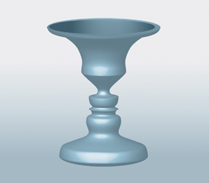
Do you see a wine glass, or do you see faces?
In this lesson you will investigate the following focusing question:
- What information about the environment do the senses of touch, smell, and taste communicate to a person’s nervous system in order to maintain homeostasis?
 Module 1: Lesson 4 Assignment
Download a copy of the Module 1: Lesson 4 Assignment to your computer now. This assignment is comprised of a lab where you will have the opportunity to choose between the senses of touch, taste, or smell. You will design your own lab to examine the characteristics of one of these senses. You will receive further instructions about how to complete this assignment later in the lesson.
You must decide what to do with the questions referred to in this lesson that are not marked by the teacher. There are also additional questions in the textbook that you may choose to complete to support your learning. Discuss your work with your teacher at any time.
Remember that these questions provide you with the practice and feedback that you need to successfully complete this course. You should respond to all of the questions and place your answers in your course folder.
You may choose to summarize the information from this lesson in notes, concept organizers, charts, diagrams, or podcasts. Store your work in your course folder.
As you work through Lesson 4, continue to work on your Module Assessment. Research the relationships between Alzheimer’s disease and interruptions in a person’s ability to interpret taste, smell, touch, and temperature. Store any relevant findings in your course folder.
Before you begin Lessons 4 to 6, you may wish to read Chapter 12, on pages 404 to 433 in your textbook, for an overview of the material these lessons will cover.
1.20. Page 2
Module 1—The Nervous System
 Explore
 Read
Read
© tillydesign/shutterstock
You can undoubtedly name the five basic senses that allow you to gather information about your environment—sight, smell, touch, hearing, and taste. To begin this lesson, read pages 406 to 409 in the textbook to explore the special type of cells, sensory receptors, that respond to stimulation from the environment. You may choose to summarize the information from your reading in notes, a concept organizer, a chart, a diagram, or a podcast. Store your work in your course folder.
The senses are classified by the type of energy that stimulates the sensory receptors. You will discover how each sense has unique receptors for detecting changes. Types of sensory receptors include photoreceptors, mechanoreceptors, chemoreceptors, osmoreceptors, and thermoreceptors. These special nerve endings—or specialized nerve cells—convert the energy stimulus into electrochemical energy, a nerve impulse. You will study the electrochemical transmission of nerve impulses in Lesson 7 of this module.
senses: specialized mechanisms or functions by which an organism is receptive and responsive to a certain class of stimuli, which are typically external (as in the senses of sight, hearing, touch, and pain) but also may be internal (as in sensing the temperature of the blood or the levels of carbon dioxide)
sensory receptor: a cell or a group of cells that is specialized to receive stimuli that provide information about the body’s external conditions (through sight, hearing, taste, smell, or touch) and internal conditions (such as temperature, pH, glucose levels, and blood pressure)
photoreceptor: a sensory receptor that responds to light stimuli, allowing people to see images and colours
mechanoreceptor: a sensory receptor that detects physical deformations in the body’s environment associated with pressure, touch, stretch, motion, and sound
chemoreceptor: a sensory receptor that transmits information about the solute concentration in a solution or about individual kinds of molecules in solution
osmoreceptor: a sensory receptor that detects changes in osmotic pressure, pressure due to water movement
thermoreceptor: a sensory receptor that detects heat or cold
sensation: the reception and processing by the brain of a nerve impulse sent by an activated sensory receptor
perception: the interpretation of sensory information by the cerebral cortex
These changes, called sensations, are communicated to specific areas of your brain, including the occipital, temporal, parietal, or frontal lobes of the cerebrum; the hypothalamus; and the cerebellum. Your brain and your friend’s brain don’t interpret information the same way. Your interpretations are based on your unique experiences. These differing interpretations result in different perceptions.
 Try This
© Glenn Frank/iStockphoto
TR 1. Prepare three bowls of water—one with ice water, one with water at room temperature, and one with water as hot as you would prepare it for a bath. Put one hand in the ice water for several minutes; then put it into the water that is at room temperature. How does the water at room temperature feel in comparison to the ice water? Put the other hand into the bowl with the hot water for several minutes. Now put this hand into the water at room temperature. How does the water at room temperature feel in comparison to the hot water, and how does it compare to what you felt after putting your hand in the ice water? Did the temperature feel different? Was your perception of the water at room temperature different after the immersion of your hand in the ice water and the hot water?
After holding your hand in ice water for several minutes, the pain of the cold does not seem so excruciating. If the sensory receptors are repeatedly stimulated, sensory adaptation occurs—this is why factory workers no longer notice the hum of machinery after working for a period of time. If you worked at a feedlot, do you think you would get used to the smell of the animals and their manure to the point where you would no longer notice the odour? Sometimes the brain perceives information differently from the sensory information it receives. This is called an illusion. Look at “Figure 12.4” on page 408 of your textbook for examples of optical illusions.
1.21. Page 3
Module 1—The Nervous System
Taste
 Read
Read

© Monkey Business Images/shutterstock
To understand the sensation of taste, read “Taste” on pages 425 and 426 in your textbook. You may wish to make summary notes and support your learning with diagrams. Store this information in your course folder for reference.
taste bud: a sensory organ composed of a taste pore, taste cells, and sensory fibres of a sensory neuron involved in initiating taste sensations
When you smell the aroma of pizza, you may start to salivate. This means the aroma, or gases, from the pizza are dissolved in your mouth by saliva. Taste buds on the surface of the tongue can only detect a taste when chemicals, such as these gases, are dissolved on the tongue. The taste buds, or chemoreceptors, then send an impulse to the brain. Chemoreceptors detect four basic tastes—salty, sweet, sour, and bitter. “Figure 12.25” on page 426 of the textbook will give you a general idea of where various taste receptors are located on your tongue. To test it yourself, try resting a sour candy on various parts of your tongue.
Your taste likes and dislikes appear to have a homeostatic value. They may be an indicator of what the body needs to retain or they may help to restore homeostasis. For example, a liking for sugar and salt helps satisfy the body’s need for carbohydrates, minerals, and some amino acids. Because many poisons and spoiled foods are bitter, a person’s dislike of bitter food is an instinctive, protective response.
 Self-Check
SC 1. What four basic tastes do most scientists agree are sensed by most people? Name at least one food that illustrates each taste.
SC 2. Supertasters are people whose sense of taste is significantly more sensitive than the average person's. Hypothesize how the anatomy of taste reception may differ in a supertaster as compared to a person with an average sense of taste.
SC 3. Explain why the taste bud is considered a “sense organ.”
SC 4. Hypothesize whether the taste buds that are sensitive to saltiness are the same taste buds that are sensitive to bitterness or sweetness.
SC 5. Draw a flow chart to illustrate the steps from taste reception to interpretation.
Self-Check Answers
SC 1. Most people are able to taste salty, sour, sweet, and bitter.
- A salty taste is mostly due to sodium ions, as in table salt. Calcium ions can also produce the salty taste.
- A sour taste is due mainly to the presence of hydrogen ions as in most acidic foods, including oranges, lemons, and tomatoes.
- A sweet taste is perceived when sugars and some proteins are present.
- A bitter taste is characteristic of coffee, unsweetened chocolate, beer, uncured olives, tonic water, and aspirin.
SC 2. Supertasters may have more papillae, more taste buds, or more taste cells in the taste buds than the average person.
SC 3. A taste bud is considered to be a sense organ because it is composed of several types of cells, such as the taste cell with its modified ending of hairs, and the sensory neuron, which is depressed into a pore where it captures chemicals from foods. The cells work together to initiate the nerve impulse.
SC 4. Taste buds sensitive to saltiness may be different than those sensitive to sweetness or bitterness because they may have different receptors on the hair portion of the taste cell that different chemicals fit into.
SC 5. Chemoreceptors in taste buds detect chemicals in food that trigger sensory neurons to communicate nerve impulses to the brain. There, the parietal lobe, which is located in the cerebrum, interprets these impulses as a sensation.
1.22. Page 4
Module 1—The Nervous System
Smell
 Read
Read

© Terrie L. Zeller/shutterstock
How does scent or fragrance affect you? For example, what happens when you catch the scent of a fragrance as you approach somebody? That smell may make you want to get closer, but it may have the opposite effect on the friend you are with. In fact, he is sneezing and his nose and eyes are running. Are your senses communicating perceptions that are different than those of your friend? To learn more about smell and what it communicates, read “Smell” on page 426 in your textbook.
Smell, much like taste, detects chemicals in solution. Olfactory receptor cells are stimulated by chemicals only when in solution. A sensory nerve communicates this information to the frontal and temporal lobes of the brain. Cells in the olfactory epithelium produce a continuous secretion of mucous in which molecules of airborne chemicals dissolve. Mucous is continuously secreted to flush away old odour chemicals so that new odour chemicals may gain access to the receptors. Smells associated with dangers, such as smoke, natural gas, or skunk, trigger the sympathetic nervous system. The perception of unpleasant odours can trigger sneezing or a gag reflex. Because your perceptions are based on your unique experiences, your perceptions can be very different from your friend’s perceptions.
olfactory (receptor) cell: a neuron located in the olfactory epithelium that is specialized to receive chemical stimuli and to initiate a nerve impulse
olfactory epithelium: a patch of tissue located in the upper part of the nasal cavity that contains mucous-secreting cells and olfactory cells, or smell cells, that can detect different smells; a sense organ for smell
 Self-Check
SC 6. Provide names for structures 1 to 14.

Inquiry into Biology (Whitby, ON: McGraw-Hill Ryerson, 2007), 427, fig. 12.26. Reproduced by permission.
SC 7. What has to happen to airborne chemicals, like those of a rose, before they can be smelled?
SC 8. What is the function of structure 10?
SC 9. What is the purpose of the olfactory cilia on the olfactory cell (receptor cell)?
SC 10. Where is the nerve impulse initiated?
SC 11. Outline the specific sensory pathway involved in smelling a rose.
Self-Check Answers
SC 6.
Structure 1: frontal lobe of cerebrum
Structure 2: olfactory bulb on underside of cerebrum
Structure 3: olfactory epithelium
Structure 4: airborne chemicals from rose
Structure 5: nasal cavity
Structure 6: olfactory bulb enlarged
Structure 7: sensory neuron
Structure 8: bone
Structure 9: sensory fibres
Structure 10: olfactory gland
Structure 11: olfactory (smell) cell
Structure 12: cilia (hairs) on olfactory cell
Structure 13: odour molecules dissolved in mucous
Structure 14: olfactory epithelium
SC 7. Airborne chemicals have to be dissolved before they can fit into receptors on olfactory cells.
SC 8. The olfactory glands (structure 10) continually produce mucous to dissolve airborne chemicals and to wash away old odours.
SC 9. The cilia (hairs) increase the surface area so that more odour molecules may reach more receptors.
SC 10. The nerve impulse is initiated at the cilia on the olfactory cell and transmitted to the neurons in the olfactory bulb.
SC 11. The main steps of smell perception include the following:
- Odour molecules are breathed into the nasal cavity.
- Odour molecules dissolve in mucous produced by olfactory glands of the nasal epithelium.
- The molecules in solution attach to receptors on the cilia (hairs) of olfactory cells.
- The sensory receptors or olfactory cells (specialized neurons) initiate nerve impulses that are sent via the sensory neuron to the olfactory bulb and then to the temporal lobe of the cerebrum where it is interpreted as a scent.
1.23. Page 5
Module 1—The Nervous System
Touch
 Read
Read
You are about to be introduced to a new student in your school. The room is so hot, and your hand is so sweaty that it feels sticky. When you reach out to shake hands, your realize that this girl has a firm handshake. Your brain tells you that the finger you sprained in basketball practice is hurting from the pressure of the handshake. You wince in pain and pull away quickly. To understand more about touch, read pages 427 to 429 in the textbook.
The mechanoreceptors for touch are located all over the body, but they aren’t evenly distributed. Consider how sensitive your fingertips and lips are in comparison to the back of your hand. Your fingertips and lips can detect light touch, pressure, pain, and high and low temperatures. In the previous activity, when you put your hand in ice water and held it there, it wasn’t long before you felt the freezing sensation. How would it feel if you put your elbow in the ice water? Do you think you could keep your elbow in the ice water longer than your hand?
Sensory Receptors in the Skin

Inquiry into Biology (Whitby, ON: McGraw-Hill Ryerson, 2007), 429, fig. 12.27. Reproduced by permission.
In the graphic above, the structure labelled “free nerve endings” is a free dendrite ending found in thermoreceptors (temperature receptors) and some pain receptors. The remaining structures have enclosed dendrites in capsules. These sensory receptors are mechanoreceptors that sense touch, pressure, vibration (on and off pressure), and stretch stimuli. The Krause end bulbs, shown on the left, and Meissner corpuscle, shown at the top right, are sensitive to light touch but are located in different parts of the body.
From the diagram, can you hypothesize which type of receptor is more concentrated in your hand than in your elbow? Does this explain why your hand was more sensitive to the cold water than your elbow was?
 Try This
TR 2. Bend a paper clip into a U-shape with the two ends about 2 mm apart. Close your eyes, and gently push the open ends of the paper clip into the palm of your hand. Then, gently press the paper clip against your shoulder and your elbow. In which location could you distinguish the two prongs as being separate?
What do your results suggest about the number of mechanoreceptors for touch in the palm of your hand in comparison to the number in your shoulder? You might want to look at “Figure 12.27” on page 429 in the textbook to review the various types of touch receptors.
 Self-Check
SC 12. Complete this receptor table.
 Module 1: Lesson 4 Assignment
You will complete a lab as part of your Lesson 4 Assignment.
 Lab
Lab
In this lab you will develop your own experimental design to study one of the senses. You may choose to study taste, smell, touch, or temperature.
Retrieve the copy of the Module 1: Lesson 4 Assignment that you saved to your computer earlier in this lesson. Complete the assignment. Save your completed assignment in your course folder. You will receive instructions about when to submit your assignment to your teacher later in this lesson.
1.24. Page 6
Module 1—The Nervous System
 Reflect and Connect
Complete one of the two scenarios that follow.
Scenario 1
The sensations of smell and taste are closely related. In Biology 20 you learned about the respiratory system. Based on your knowledge of physiology, reflect on why is it more difficult to taste or smell food when you have a cold?
Prepare a concept map, a diagram, or a podcast to illustrate how Lesson 4 fits with the previous three lessons. Remember to link the sensory receptors to the appropriate parts of the central or peripheral nervous system, and include the specific parts of the brain that are involved.
Scenario 2
In this lesson you were asked to think about sensing the smell of pizza or a person’s fragrance, the taste of pizza, the temperature of the room, or the pain in your hand. Write a poem, make a comic strip, or draw diagrams to illustrate the communication and processing of one of these sensations. Include in your example the stimulus, the type of sensory receptor, the sensory neuron, and the part of the brain that are involved in the process of sensation.
Share your work for discussion with your peers and your teacher.
 Self-Check
SC 13. Identify the statement that best compares sensation to perception.
- A sensation occurs when nerve impulses from the sense organs reach the cerebral cortex; perception is an interpretation by the brain about the meaning of sensations.
- A sensation occurs when nerve impulses from the sense organs reach the spinal cord; perception is an interpretation by the spinal cord of the meaning of sensations.
- A sensation occurs when nerve impulses from the sense organ bypass the spinal cord and go directly to the brain; perception is an interpretation by the sensory receptors of the meaning of sensations.
- A sensation occurs when nerve impulses from the sense organs reach the cerebral cortex; perception is an interpretation by the peripheral nervous system of the meaning of sensations.
SC 14. You walk into a pizza restaurant and immediately notice the smells associated with cooking pizza. After a few minutes, you no longer notice these odours. This phenomenon can be explained by a process called
- sensory accommodation
- a reflex arc
- sensory adaptation
- sensory perception
SC 15. Which of the following is an INCORRECT match between the sensory receptor and the stimulus that it responds to?
- photoreceptor – light energy
- chemoreceptor – pressure
- mechanoreceptor – sound waves
- thermoreceptor – change in radiant energy
SC 16. The olfactory receptors in a dog’s nose would be classified as
- photoreceptors
- mechanoreceptors
- chemoreceptors
- proprioceptors
SC 17. Which of the following structures would you NOT mention if you were tracing the path involved in tasting something sweet?
- taste bud
- olfactory receptor cell
- sensory neuron
- parietal lobe of cerebrum
Use the following information to answer questions SC 18 and SC 19.
| The tips of the fingers are sensitive enough to discriminate raised points on a surface, as well as the locations of these points. Knowing this, in the nineteenth century Louis Braille invented the Braille system of reading for people who are visually impaired. Each letter of the alphabet is represented by up to six raised dots. A visually impaired person who has learned Braille can read up to 50 words a minute. |
SC 18. Does a person preparing Braille script need to know if the visually impaired reader is left-handed or right-handed and, thus, change the order of the script accordingly?
- No, because the sensations from both the right hand and the left hand are carried by the spinal cord into the same hemisphere of the cerebrum.
- No, because the sensations received separately by the left and right cerebral hemispheres from the right and left hands are integrated before they are interpreted into language.
- Yes, because the sensations from the right and left hands are carried to the left and right cerebral hemispheres, respectively, and therefore the information is interpreted in reverse order.
- Yes, because the movements of the right and left hands are initiated by opposite sides of the cerebral cortex.
SC 19. This sentence can be read using the eyes, but it can also be read using the fingertips if it is printed in Braille. This fact illustrates that
- the brain can form the same meaning from different sensations
- nerve impulses initiated by touch are identical to nerve impulses initiated by sight
- receptors for touch and receptors for sight respond to the same environmental stimuli
- the lobe of the brain that receives sensory stimuli from the eyes is the same lobe that receives sensory stimuli from the fingers
Self-Check Answers
SC 13. C
SC 14. C
SC 15. B
SC 16. C
SC 17. B
SC 18. B
SC 19. A
Reflect on the Big Picture
Lesson 4 focused on how you gather information about your environment through the senses of taste, smell, and touch. In the Big Picture, you gathered information about the sound of laughter, the pain of a handshake, and the temperature of a room just by being there. This information had to be received by your sensory receptors and communicated through sensory nerve impulses to different lobes of the cerebrum, such as the temporal and occipital lobes.
The brain interprets this information and causes your body to feel something or to do something in response. If a fragrance is pleasant, you may move closer to the person wearing it, inhale more deeply, and feel pleasure. But, do you remember the sneezing response that your friend had to the odours? Using the information from this lesson, reflect on these possible responses to a stimulus. Store this response in your course folder.
 Going Beyond
If you would like to further explore the senses of taste and touch beyond the information presented in your textbook, watch the video “Science of the Senses—Taste and Touch” at CBC.ca/Nature of Things. To locate this video, use search terms such as “CBC,” “Nature of Things,” and “Senses.”
 Module 1: Lesson 4 Assignment
Submit your completed Module 1: Lesson 4 Assignment to your teacher for assessment. Share your response to the Reflect and Connect activity on the discussion board. After sharing your work with your peers and teacher, store your response to the Reflect and Connect activity in your course folder.
1.25. Page 7
Module 1—The Nervous System
 Lesson Summary
In this lesson you investigated this focusing question:
- What information about the environment do the senses of touch, smell, and taste communicate to a person’s nervous system in order to maintain homeostasis?
You discovered how the body is able to gather information about the external and internal environment in order to maintain a constant internal environment, or homeostasis. The senses are the functional categories that scientists use to classify how the body gathers information. You studied the specialized neurons that respond to stimuli (types of energy). These specialized nerve cells are identified as
- mechanoreceptors
- chemoreceptors
- thermoreceptors
- photoreceptors
The sensory receptors for taste and smell are chemoreceptors, while touch receptors are mechanoreceptors. Temperature sensory receptors are thermoreceptors. The sensory receptors are able to initiate a nerve impulse that is transmitted through a sensory neuron to a specific part of the brain that is able to interpret the information. The brain initiates a response that maintains or returns the body to homeostasis.
Lesson Glossary
Consult the glossary in the textbook for other definitions that you may need to complete your work.
chemoreceptor: a sensory receptor that transmits information about the solute concentration in a solution or about individual kinds of molecules in solution
mechanoreceptor: a sensory receptor that detects physical deformations in the body’s environment associated with pressure, touch, stretch, motion, and sound
olfactory (receptor) cell: a neuron located in the olfactory epithelium that is specialized to receive chemical stimuli and to initiate a nerve impulse
olfactory epithelium: a patch of tissue located in the upper part of the nasal cavity that contains mucous-secreting cells and olfactory cells, or smell cells, that can detect different smells; a sense organ for smell
osmoreceptor: a sensory receptor that detects changes in osmotic pressure, pressure due to water movement
perception: the interpretation of sensory information by the cerebral cortex
photoreceptor: a sensory receptor that responds to light stimuli, allowing people to see images and colours
sensation: the reception and processing by the brain of a nerve impulse sent by an activated sensory receptor
senses: specialized mechanisms or functions by which an organism is receptive and responsive to a certain class of stimuli, which are typically external (as in the senses of sight, hearing, touch, and pain) but also may be internal (as in sensing the temperature of the blood or the levels of carbon dioxide)
sensory adaptation: the tendency of sensory neurons to become less sensitive when they are repeatedly stimulated
sensory receptor: a cell or a group of cells that is specialized to receive stimuli that provide information about the body’s external conditions (through sight, hearing, taste, smell, or touch) and internal conditions (such as temperature, pH, glucose levels, and blood pressure)
taste bud: a sensory organ composed of a taste pore, taste cells, and sensory fibres of a sensory neuron involved in initiating taste sensations
thermoreceptor: a sensory receptor that detects heat or cold
1.26. Lesson 5
Module 1—The Nervous System
Lesson 5—Photoreception—the Eye
 Get Focused
When you are in a room full of people, you are constantly gathering information about those around you. You respond to the sights, smells, sounds, and feel of the room. Your eyes are one of the best sensory tools you can utilize to gather information from your external environment. In fact, 80% to 90% of all sensory information is received by your eyes. Consider this fact the next time you find yourself trying to make sense of a new environment or situation.
Your ability to interpret the visual information you receive is a complex process that is dependent on the physiology of the eye and a complex series of reactions that are not entirely understood. In Lesson 2 you studied the parts of the brain, so you should now be capable of making the connection between how a nerve impulse is transmitted to the occipital lobe of the cerebrum and how the image is then interpreted by the same part of the brain. The work you did in Lesson 4 will help you understand how perception results from the cerebral cortex interpreting the meaning of the sensory information received.
How much do you remember about what you saw after you leave a room full of people? If you were to compare your perceptions with another person who was in the same room, do you think your visual memories would be the same? For an interesting example of how what you see is perceived, observe the rotating dancer.

According to your perception, in which direction is she rotating? Were you able to change the direction of her rotation? Your perception is based on how your brain interprets visual information. In this lesson you will learn about eye function and the communication of visual information. You will also see how these processes influence your perceptions of what you see.
In this lesson you will explore the following questions:
- What are the major parts of the eye?
- How do these parts function?
- How do these parts work together to communicate with the nervous system in order to facilitate the integrated act of seeing?
 Module 1: Lesson 5 Assignment
Your teacher-marked Module 1: Lesson 5 Assignment requires that you complete and submit the following:
- an eye lab
- a table of vision defects and disorders
Download a copy of the Module 1: Lesson 5 Assignment to your computer now. You will receive further instructions about how to complete this assignment later in the lesson.
You must decide what to do with the questions that are not marked by your teacher.
Remember that these questions provide you with the practice and feedback that you need to successfully complete this course. You should respond to all of the questions and place those answers in your course folder.
There are also many valuable practice questions in your textbook that you can use to evaluate your understanding. You can ask your teacher for direction on choosing extra questions or for assistance with your answers.
Any work completed in summary notes, results of Self-Checks, concept maps, flow charts, or diagrams that you develop should be stored in your course folder as reference for future study. You will also save a discussion board activity about the risks and benefits of laser surgery.
The Module Assessment for Module 1 involves a study of Alzheimer’s disease. As you work through this lesson examining the structures and functions of the eye, consider whether there are any links to the symptoms of Alzheimer’s disease. Store your research in your course folder for use in the final preparation of your response to the Module Assessment.
This lesson may take longer than 80 minutes to complete. You should devote the time necessary to complete your understanding of the structures and functions of the eye and the features of vision because this is the most important of the senses.
Required Materials and Equipment
For this lesson you will have the choice of conducting either a virtual dissection of an eye or an actual dissection of an eye. To conduct the dissection lab of a real eye, you will need
- a preserved cow eye obtained from a biological supply house or a fresh cow eye obtained from a butcher
- rubber gloves
- safety goggles
- a clean dissecting tray or a clean foam tray
- sharp scissors with pointed tips
- a sharp knife or a scalpel
- a bleach solution to be used for cleanup
1.27. Page 2
Module 1—The Nervous System
 Explore
The Structures of the Eye
 Read
Read
refract: to bend light as it passes through a substance with a different desnsity
retina: the innermost layer of the eye that contains the photoreceptors
optic nerve: a collection of sensory neurons that carries sensory information from the photoreceptors to the brain
The eye is an amazing organ! It contains many structures that must all be functioning properly in order for you to see. Several of these structures refract, or bend, light and focus it on the different photoreceptors that make up the retina. Once the photoreceptors are stimulated by light energy, they convert it into an electrochemical nerve impulse. Sensory neurons in the optic nerve communicate this nerve impulse to interneurons in the occipital lobe of the brain, where visual information is then processed. To understand the parts of the eye and how they contribute to vision, read from page 410 to the heading “Conditions Affecting the Cornea and Lens” on page 412 of the textbook.
As you read, keep track of the vision disorders and their respective symptoms. You will need this information for an upcoming assignment in which you will be required to develop a summary chart of eight vision disorders. For each disorder, you will include information about the structure affected, the symptoms of the disorder, the physiology of the disorder, and technologies that can be used to detect or treat the disorder.
It is always an excellent idea to support your understanding with a diagram. You may choose to include diagrams in the work you will store in your course folder. If you choose to include diagrams, be sure to include brief descriptions of the functions of any structures and brief summaries of the steps of different pathways.
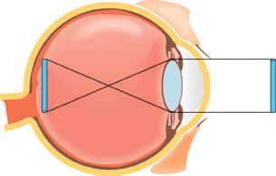
 Try This
There are three Try This activities in this lesson that can be used to verify your understanding of the parts of the eye and their functions as well as vision issues related to the dysfunction of one or more of these structures. There is a choice of two possible activities for each one. You may choose to do all of the Try This activities for a complete review of the concepts you have been studying.
TR 1. Choose one of the following activities.
Complete the eye function drag-and-drop activity to review the structures and functions of the eye.
OR
Watch the “Bio Review: Eye Structure and Function” section of “The Eye: Vision and Perception: A Whole World to See” that deals with the structures of the eye. You may be required to enter a username and password in order to access this video. Contact your teacher for this information.
After watching the video, make a diagram of the eye that includes summary notes about the function of each part of the eye. Store this work in your course folder for review purposes.
Accommodation
ciliary muscle: a ring of muscle behind the iris that is attached to the lens by suspensory ligaments and is involved in changing the shape of the lens
accommodation: the process of changing the shape of the lens from round and fat to thin and flat, and vice versa, so that light can be focused on the retina to accommodate vision of objects near and far away
The ciliary muscle and suspensory ligaments are excellent examples of how the sympathetic and parasympathetic nervous systems work together to establish homeostasis in vision. By changing the shape of the lens, light rays are bent differently, allowing you to focus on the words of this sentence or on the words on a poster across the room. This process is called accommodation.
Try This
TR 2. Choose one of the following activities.
Put the following lists in the correct order. You may wish to look at “Figure 12.10” on page 412 of the textbook to help you figure out the correct order of events. This activity is similar to the numerical-response questions that you will complete on the Diploma Exam. Note that the number assigned to the event does not imply the correct order. In this type of numerical response, you take the assigned number of the event and place that number in the correct order or sequence in the response area.
- Sympathetic Nervous System
- tension pulls on lens
- focus on far objects
- lens flattens
- tension on suspensory ligament
- ciliary muscle relaxes
Place the numbered events in the order that illustrates the correct order or sequence in which these events occur.
___, ___, ___, ___, ___
- Parasympathetic Nervous System
- ciliary muscle contracts
- tension on suspensory ligament relaxes
- less pull on lens
- lens bulges
- focus on near objects
Place the numbered list in the correct order.
___, ___, ___, ___, ___
OR
Review the accommodation reflex by watching “Bio Review: Accomodation Reflex” segment of “The Eye: Vision and Perception: A Whole World to See.”
You may be required to enter a username and password to be able to view the video. Contact your teacher for this information.
Try This Answers
TR 2.
- The correct numerical sequence of events is 5, 4, 1, 3, 2.
- The correct numerical sequence of events is 1, 4, 3, 2, 5.
The Lens
Read
The flexibility of the lenses decreases as people age. As a result, many people must wear reading glasses to focus the details of nearby objects. Hyperopia is the term used to describe the ability to see distant objects clearly and the inability to focus on objects that are close. You likely know the more common term for this condition—farsightedness. Myopia is the condition of being able to clearly see objects that are close but not being able to focus on objects that are farther away. The common term for myopia is nearsightedness.
To review what enables the lens to change its shape in order to successfully focus an image, you may wish to review pages 412 and 413 in the textbook. When you have finished the reading, make summary notes about the concepts you learned. You may also wish to draw a labelled diagram. Store your information in your course folder for review purposes.
Cataracts are another condition that affects the lens. Astigmatism is a condition that affects the cornea. Both these conditions distort vision. Remember to add them to your table of disorders. You may choose to research these conditions and add the information about these disorders to your table now. At the end of this lesson, you will submit your table to your teacher for assessment.
To develop your mastery of the concepts you have been reading about, do questions 1, 2, 3, and 5 on page 418 of your textbook. You may discuss your responses with your teacher if you wish.
hyperopia: farsightedness, or the inability to focus objects that are close, caused by an eyeball that is too short, which causes light to be focused behind the retina
myopia: nearsightedness, or the inability to focus objects that are far away, caused by an eyeball that is elongated, which causes light to be focused in front of retina rather than directly on it
cataract: a cloudy or grey-white area on the lens caused by deterioration of the protein composing the lens
Cataracts prevent the passing of light to the photoreceptors of the retina.
astigmatism: an uneven curvature of the cornea or lens, resulting in uneven focusing, which results in poor vision
cornea: a transparent portion of the sclera (located at the front of the eye) that allows light to enter the eye and, in the process, refracts or bends the light rays so that they can be focused on the retina
The Retina and Supporting Structures

© Tim Mainiero/shutterstock
The inner layer of the eye consists of the delicate retina, which contains two different types of photoreceptors called rods and cones. Directly in line with the middle of the lens is an area of the retina called the fovea centralis, or fovea. This small, depressed area is where the cones are most highly concentrated. Cones are the photoreceptors that are sensitive to different colours, whereas rods are sensitive to light intensity. Rods are more spread out on the periphery of the retina. The neural fibres from the retina form the optic nerve, which is the sensory pathway between the eye and the brain. The spot where the fibres come together is the blind spot. This area is called the blind spot because it lacks receptor cells. Therefore, any light or image falling on this area goes undetected.
Organisms that see well at night, like deer and cats, have a reflective layer, similar to spray paint, on the surface of the choroid layer. This structure is called the tapetum. The tapetum increases the animal’s sensitivity to low levels of light and helps these animals see at night. This layer also causes the eyes of these animals to appear iridescent or reflective in the dark.
In front of the lens is the anterior chamber, which is filled with aqueous humour. This transparent, watery fluid is produced by the ciliary body. If the aqueous humour is not successfully drained from the anterior chamber, its buildup will eventually result in the eye disorder called glaucoma. Behind the lens is the posterior chamber filled with vitreous humour—a clear to amber-coloured gel-like fluid. Both the aqueous and vitreous humours will aid the refraction of light to a small degree.
rods: one of two types of photoreceptors in the retina of the eye that is sensitive to light intensity and detect movement
Rods do not distinguish colour.
cones: one of two types of photoreceptors in the retina of the eye that is sensitive to different wavelengths of light and are, thus, responsible for distinguishing colour
There are three types of cones: one sensitive to red light, one sensitive to blue light, and one sensitive to green light. The cones are responsible for acute vision, or distinguishing detail.
fovea centralis: an area of the retina that is located directly behind the centre of the lens and has a very high concentration of cones, which makes this part of the eye responsible for great visual acuity
blind spot: the area at the back of the eyeball that is deficient in rods and cones; the area where the sensory fibres come together to form the optic nerve
choroid: the middle layer of the eyeball that lies between the sclera and retina and is highly vascular and heavily pigmented
The choroid absorbs stray light rays not detected by the photoreceptors of the retina.
tapetum: a layer in the choroid that increases the absorption of light to stimulate photoreceptors in dim conditions
anterior chamber: the space in front of the iris and behind the cornea that is filled with aqueous humour
aqueous humour: a clear, watery fluid in the anterior chamber of the eye that maintains the shape of the cornea and provides oxygen and nutrients for the surrounding cells, including those of the lens and the cornea
glaucoma: a disorder caused by the malfunction of ducts that drain excess aqueous humour from the anterior chamber
The resulting pressure created by excess aqueous humour ruptures delicate blood vessels in the eye and causes deterioration of cells in the eye due to lack of nutrients. This can result in blindness if left untreated.
vitreous humour: the transparent, amber-coloured, jelly-like fluid in the posterior chamber of the eye that helps to maintain the shape of the eyeball
Try This
TR 3. Choose one of the following activities. Whichever one you choose, be sure to take summary notes and then save them in your course folder.
Review the parts and functions of the eye by reading pages 414 and 415 in your textbook.
OR
Watch the following segments of “The Eye: Vision and Perception: A Whole World to See”
- “Bio Probe: Cow Eye Dissection”
- “Bio Review: Eye Structure and Function”
You may be required to enter a username and password to be able to view the video. Contact your teacher for this information.
1.28. Page 3
Module 1—The Nervous System
 Lab—Dissection of the Eye
For this dissection lab, you have the choice of either conducting your own hands-on dissection of a real cow eye or participating in a virtual dissection of an eye. If you wish, you may do both labs.
If you choose to do the hands-on lab, you will need access to either a preserved cow eye from a biological supply house or a fresh cow eye from a butcher. A cow eye is typically used for this dissection as its anatomy is very similar to that of the human eye.
Review the list of required materials for this lab:
- a preserved cow eye obtained from a biological supply house or a fresh cow eye obtained from a butcher
- rubber gloves
- safety goggles
- a clean dissecting tray or a clean foam tray
- sharp scissors with pointed tips
- a sharp knife or a scalpel
- a bleach solution to be used for cleanup
To complete the hands-on dissection, refer to “Investigation 12.A: Dissection of an Eye” on page 417 of your textbook. You may also find it helpful to view the virtual dissection to help you through the procedure.
To view the virtual dissection, watch the "Bio Probe: Cow Eye Dissection" segment of “The Eye: Vision and Perception: A Whole World to See.” You may be required to enter a username and password to be able to view the video. Contact your teacher for this information.
As you conduct either dissection, remember that the retina is composed of two important types of photoreceptors called rods and cones. Try to locate the fovea centralis, the small depression in the retina. This area is specialized for sensitivity to colour and acute images as it has a high density of cones. The rods, which are sensitive to light intensity, cover the surrounding area of the retina.
The tapetum is a specialized layer located in the choroid. This structure is only typical of animals, such as the cow, that are able to see well in the dark. Try to find the tapetum.
The role of the aqueous and vitreous humours should become obvious in the dissection. Think about glaucoma—the disorder that results when the aqueous humour is not successfully drained from the anterior chamber. Be sure to add this disorder to your list.
Regardless of which dissection you complete, make sure you create a diagram of the eye that includes the following structures:
- sclera
- choroid
- retina
- cornea
- iris
- pupil
- ciliary muscle
- suspensory ligaments
- lens
- optic nerve
- fovea centralis
- optic disc or blind spot
- aqueous humour
- vitreous humour
You may choose to add these to a diagram you have already drawn or develop a new diagram based on the findings of the dissection.
1.29. Page 4
Module 1—The Nervous System
 Module 1: Lesson 5 Assignment
Retrieve the copy of the Module 1: Lesson 5 Assignment that you saved to your computer earlier in this lesson. Complete Part 1—Lab, which is based on the dissection of the eye. Save your completed assignment in your course folder. You will receive instructions about when to submit your assignment to your teacher later in this lesson.
 Self-Check
SC 1. Complete the following crossword puzzle to consolidate your understanding of the different structures of the eye and their functions.
Self-Check Answers
SC 1.
Across
1. sclera
3. choroid
7. blind spot
8. pupil
9. ciliary muscle
11. iris
12. tapetum
13. lens
14. vitreous humour
15. fovea centralis
16. optic nerve
Down
2. conjunctiva
4. rods
5. ciliary body
6. cornea
10. cones
The Retina
 Read
Read
You should now be familiar with the different structures of the eye and have a good understanding of each structure’s role in vision. You should understand the role of the cornea and lens in bending and focusing light onto the retina. The retina is the key structure by which light energy is converted into an electrochemical impulse. This sensory impulse is then communicated to the brain via the optic nerve. When it reaches the occipital lobe of the brain, the information that was transmitted as an impulse is processed by interneurons. To further understand how the retina converts light energy into electrochemical impulses, read pages 414 to 416 in the textbook.
Study the diagram below to locate the four layers of cells in the retina:
- pigmented cells: closest to the choroid, specialized to form the tapetum in some animals
- rods and cones: the actual photoreceptors, located above the pigmented layer
You might expect the photoreceptors to be in the direct path of incoming light, but the rods and cones are further covered by layers of transparent neurons.
- bipolar cells: activated by rods and cones
- ganglion cells: closest to the vitreous humour
Roll your mouse over the labels on the diagram of the retina to highlight the different structures.
ganglion cells: special sensory neurons that communicate with bipolar cells in the retina to transmit a nerve impulse to the brain
These cells have long axons that converge at the back of the eye to form the optic nerve.
Now that you have located the different layers, note the direction of the light striking the retina. Light travels through the transparent layer and strikes the rods and cones. These photoreceptors are then prompted to stop emitting an inhibiting neurotransmitter substance, which you will learn about in Lesson 8. The rods and cones can then communicate an electrochemical message, first to the bipolar cells and then to the ganglion cells. Axons of all ganglion cells of the retina converge at the back of the retina to form the optic nerve, which carries impulses to the brain.
Rods and Cones
When looking at the diagram of the retina, you may have noticed that rods are the most abundant photoreceptors in the retina. They are necessary for distinguishing shades of black and white as well as for distinguishing movement. Cones respond to specific wavelengths of the visible spectrum, allowing people to see colour. Cones are also needed for acute vision, as they are capable of finer discrimination of detail in bright light than are rods.
Based on your new knowledge of rods and cones, could you develop a hypothesis explaining why the eyes of birds that roost at night have only cones, whereas the eyes of owls and bats that are active at night have only rods?
You have also read about colourblindness. There are three types of cones: red sensitive, green sensitive, and blue sensitive. One reason that people can see so many variations of colour is that the sensitivity ranges of these receptors overlap. For example, yellow light stimulates both red and green cone cells; but if the red cones are stimulated more strongly than the green cones, you see orange instead of yellow. Look at “Figure 12.14” on page 414 of your textbook. If you see a number 8, you have the full range of colour vision. If you see a number 3, you have red-green colour blindness. Can you explain the varying degrees and types of colourblindness in terms of the types of cones? Colourblindness is a genetically inherited trait that will be studied in more detail in Unit C.
 Try This
TR 4. Choose one of the following activities.
To review the concepts of vision, watch the following segments of the video “The Eye: Vision and Perception: A Whole World to See.”
- “The Retina”
- “Bio Bit: Colour Blindness”
You may be required to enter a username and password to access the video. Contact your teacher for this information.
OR
Do an Internet search using search terms such as “colourblindness,” “colourblindness test,” and “Ishihara test.”
1.30. Page 5
Module 1—The Nervous System
Rods and Cones Initiate Nerve Impulses

Inquiry into Biology (Whitby, ON: McGraw-Hill Ryerson, 2007), 415, fig. 12.15. Reproduced by permission.
Rods and cones contain pigments called photopsins, which absorb light and start chemical changes. Cones contain three different pigments called iodopsins. Rods have only one type of pigment called rhodopsin. When rhodopsin absorbs light, it is bleached and breaks down into opsin, a colourless protein, and a derivative of Vitamin A called retinal. This chemical change in the rods initiates a nerve impulse in the bipolar cells, which transfers the impulse to the ganglions. The ganglions, which make up the optic nerve, then transmit the impulse to the occipital lobe of the cerebrum to be processed.
Your eye then restores opsin and retinal to rhodopsin, but this takes energy. As you will recall from Biology 20, this energy is in the form of ATP. This reverse reaction also requires Vitamin A. Knowing this, can you explain why your eyes get strained in poor lighting, why night blindness is caused by Vitamin A deficiency, and why you should eat your carrots?
iodopsin: the general name of any of the three visual pigments found in cone cells that is stimulated by light to initiate a nerve impulse
rhodopsin: a visual pigment found in rod cells that is decomposed by light into opsin and retinal
The change from rhodopsin to opsin and retinal initiates a nerve impulse.
opsin: a protein that is the result of the decomposition of rhodopsin
retinal: a derivative of Vitamin A (retinol) that is the result of the decomposition of rhodopsin and is instrumental in initiating a nerve impulse
© Gilmanshin/shutterstock
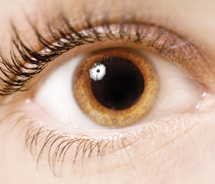
© Johanna Goodyear/shutterstock
Light and Dark Adaptation
Rhodopsin is extremely sensitive to light. Even starlight causes some molecules of rhodopsin to become bleached. In high-intensity light, rhodopsin is broken down almost as fast as it is remade. In such a situation, the rods become nonfunctional and the cones take over completely. For example, think about what happens when the lights are turned on after a movie. You are momentarily “blinded,” and all you see is white light. Your pupils constrict as a reflex to protect your retina from the bright light. Within a minute or so, the cones become sufficiently stimulated to take over once again. Over the next few minutes, visual acuity and colour vision improve. On the other hand, adaptation to low light occurs when you go from a well-lit area into a dark one. At first you see nothing but velvety darkness because your cones have stopped functioning due to lack of light, and your rods are not functioning because they have been bleached out by the bright light. Once in the dark, rhodopsin is remade and accumulates so that low-intensity light can again stimulate the rods. During adaptation to both light and dark, reflexive changes occur in the pupil.
The Pathway to the Brain
 Try This
TR 5. Did you know that each eye sees thing differently? Each eye has its own area of “blindness,” the blind spot. To find your blind spot, try the activity shown in “Figure 12.17” on page 416 of your textbook.
The information you receive from each eye is communicated through the optic nerve to the brain. Your brain processes this information into one image without gaps or blind spots. The optic nerves enter the optic chiasm on the ventral, front of the brain (see Lesson 2). In the X-shaped optic chiasma, some axons from each eye cross over to the opposite side of the brain, while others continue to their own side of the brain. The crossing over of about half of the sensory fibres ensures that each half of the occipital lobe receives the same image or part of the visual field viewed by each eye. However, keep in mind that each eye will see the image from a slightly different angle. Study the illustration provided below very carefully in order to better understand visual interpretation by the brain.
depth perception: the ability to see in three dimensions
Depth perception is possible due to the “fusing” or superimposing of the slightly different images from the two eyes by the visual cortex. This creates a three-dimensional image. Thus, by working together, the eyes provide slightly different angles of view that allow the brain to estimate distance.
The image that was reversed and inverted in the lens is at this point interpreted correctly.
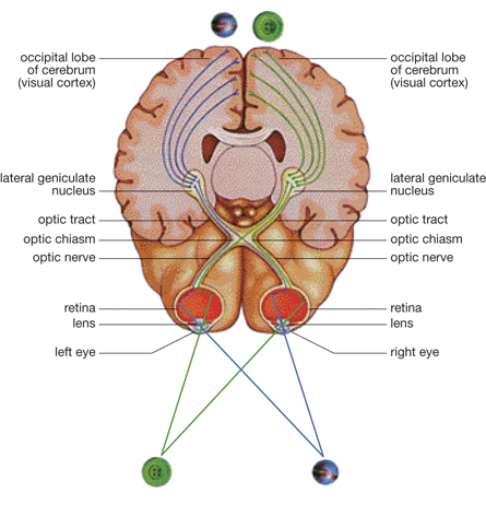
Inquiry into Biology (Whitby, ON: McGraw-Hill Ryerson, 2007), 416, fig. 12.18. Reproduced by permission.
1.31. Page 6
Module 1—The Nervous System
 Try This
TR 6. Choose one of the following activities.
Do questions 5 and 7 on page 418 of the textbook.
OR
Watch the segment called “Interpretation” in the video “The Eye: Vision and Perception: A Whole World to See,” which addresses how the brain is involved in interpretation of vision stimuli.
You may be required to enter a username and password to access the video. Contact your teacher for this information.
 Module 1: Lesson 5 Assignment
As you worked through this lesson, you learned about the different structures of the eye and their functions in facilitating vision. You were also introduced to some disorders that are the result of one or more of these structures malfunctioning.
Retrieve the copy of the Module 1: Lesson 5 Assignment that you saved to your computer earlier in this lesson. Complete Part 2—Table of Vision Defects and Disorders. Recall that you have already studied some of these vision disorders. Make sure you add these to your table. Save your completed assignment in your course folder. You will receive instructions about when to submit your assignment to your teacher later in this lesson.
 Self-Check
To ensure that you have mastered the concepts of this lesson, do this Self-Check that includes a numerical-response question and a closed written-response question. This is an excellent opportunity to practise for the types of questions that will be presented on the Diploma Exam.
Use the following information to answer question SC 2.
Visual Interpretation
The brain integrates visual information from both eyes. Some of the steps in the visual pathway are shown below. Note that these steps are NOT in the correct order and that NOT all steps are part of the visual pathway.
|
SC 2. In the four spaces below, record the assigned numbers of the four steps that are part of the visual pathway. Record the steps in the correct order in which they occur during the visual pathway.
____ ____ ____ ____
SC 3. There are two main types of light-sensitive cells (called X and Y in this question) in the healthy human retina, but they are not evenly distributed. An investigation of the distribution of sensory cell types X and Y across an area of the retina (shown as PQ on the diagram below) was developed.
The relative numbers of cell types X and Y were more or less constant, except in positions R and S. Using relative numbers, the following graph indicates the relative frequency of sensory cell type X along the axis PQ.
- Identify cell type X.
- Identify cell type Y.
- What structure is represented by region R?
- What structure is represented by region S?
- Copy the graph above and clearly superimpose a curve to indicate the relative frequency of cell type Y along the axis PQ.
SC 4. The diagram below represents the different states of the iris in bright and in dim light.
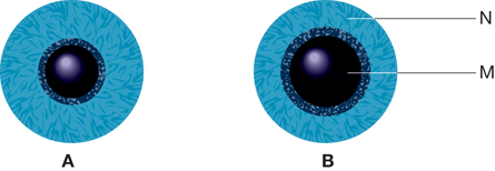
- What structures are represented by M and N?
- Briefly explain what causes the iris of eye B to have a different appearance than that of
eye A.
Self-Check Answers
SC 2. 6, 1, 4, 3
SC 3. a. i. Cell Type X represents the cones.
ii. Cell Type Y represents the rods.
iii. Region R represents the blind spot.
iv. Region S represents the fovea centralis.
b. Cell Type Y would be constant with a sharp dip in region R and S.
SC 4. i. M and N represent the circular muscle of the iris or, more specifically, M represents the circular muscle and N represents the radial muscle.
ii. In bright light, represented by A, the parasympathetic nervous system (a division of the autonomic nervous system) stimulates the contraction of the circular muscle (M) of the iris, and allows the relaxation of radial muscle (N). This reduces the pupil diameter and allows less light to enter the eyeball and strike the retina.
In dim light, represented by B, the sympathetic nervous system (a division of the autonomic nervous system) stimulates the contraction of the radial muscle (N) of the iris and allows the relaxation of circular muscle (M). This increases the pupil diameter, which allows more light to enter the eyeball and strike the retina.
1.32. Page 7
Module 1—The Nervous System
 Reflect and Connect
Most people are left-brain dominant, but some people are right-brain dominant. The dominance of one side of your brain over the other affects how you perceive what you see. Your brain is crucial in the integration, interpretation, and perception of what you see. Two people looking at the same thing may see it very differently!
Choose one of the following written-response questions. Reflect upon the question and consider what you have learned in this lesson about the eye, how it receives a stimulus, how the message is sent to the brain, and how the brain processes that information; then respond in paragraph format. Store your response in your course folder for future reference.
RC 1. Look at a picture, clock, or some distinctive object located on the other side of the room. Cover or close one eye. Concentrate on that object for a few moments. Now, cover or close the other eye. What happens to the image when you switch from viewing it first with one eye and then with the other? What does this simple experiment illustrate about vision?
RC 2. The actual visual sensation that you experience is not always an exact representation of the visual information that is picked up by your eyes. This is because the brain is susceptible to all sorts of past and present influences that modify your perception. Visual information is modified by the brain so that you see the image that your brain is conditioned to see. Do an Internet search using the term “old woman young woman perception.” Your search engine will display a number of illustrations. Follow a link to an illustration that plays on perception. Look at the illustration. What do you see? It is possible to see a young woman or an old woman depending on how you look at it. Perhaps you are able to see both. The image that you see, or your perception of the image, depends on your brain and your life experiences.
 Discuss
Would you have laser surgery to change the shape of your lens? Many nearsighted people are now considering laser surgery to eliminate the need for glasses. Research this technology and identify any possible benefits and risks associated with the procedure. Address and explain at least two benefits and two risks of the procedure. Decide whether you would have such surgery if you were nearsighted, and justify your decision.
Post your findings on the discussion board. Ask classmates to comment on whether they would have the surgery. You may prepare your findings as a chart that lists the pros and cons of the procedure, or you may record your findings in point form.
Reflect on the Big Picture
In this lesson you continued exploring the senses. Photoreception, or the sense of vision, involves the largest number of sensory receptors in the body. The eyes gather approximately 80% to 90% of the information about the external environment and transmit it to the brain.
Complete the following statements by filling in the blanks.
Statement 1: As you walked toward the group of people and focused on one person, her image was kept in focus by what reflex?
Statement 2: The _____ adjusted the amount of light entering the eye.
Statement 3: The _____ were activated to allow you to see colours.
Statement 4: The _____ integrated and interpreted this information.
Based on this information that you received and the visual perception that you formed, you decided to say hello to the new girl in your class.
Reflect on the Big Picture Answers
Statement 1: Her image was kept in focus by the accommodation reflex.
Statement 2: The papillary reflex adjusted the amount of light entering the eye.
Statement 3: The cones were activated to allow you to see colours.
Statement 4: The occipital lobes of the cerebral cortex integrated and interpreted this information.
At this point, you should research the relationship between Alzheimer’s disease and vision. Store any additional information in your course folder in preparation for your Module Assessment.
Keep the following key questions in mind when reviewing:
- What are the major structures of the eye?
- How do these structures function?
- How do they communicate with the nervous system to support the integrated act of seeing?
 Module 1: Lesson 5 Assignment
Submit your completed Module 1: Lesson 5 Assignment to your teacher for assessment.
1.33. Page 8
Module 1—The Nervous System
 Lesson Summary
In this lesson you explored the following focusing questions:
- What are the major parts of the eye?
- How do these parts function?
- How do these parts communicate with the nervous system to support the integrated act of seeing?
Through your readings, the dissection you completed, your research, and the videos you watched, you learned about the structures of the eye and their functions. By exploring the structures of the photoreceptors, called the rods and cones, you learned how a nerve impulse is initiated and transmitted to the occipital lobe of the cerebrum. It is there that the sensations are interpreted. Things like the frequency of nerve impulses, the origin of the impulse in the retina, and previous experiences and memory all factor into the interpretation of light stimuli. Not everyone will interpret these sensations in the same way, as is observed with optical illusions. In the Lesson 5 Assignment, you researched many common visual defects and disorders that affect the functioning of the eyes and impede proper vision. You also examined numerous technologies that have been devised to correct these disorders.
Lesson Glossary
Consult the glossary in the textbook for other definitions that you may need to complete your work.
accommodation: the process of changing the shape of the lens from round and fat to thin and flat, and vice versa, so that light can be focused on the retina to accommodate vision of objects near and far away
adaptation: the process by which the iris adjusts the diameter of the pupil based on light conditions, thus controlling the amount of light that enters the eye and strikes the retina
anterior chamber: the space in front of the iris and behind the cornea that is filled with aqueous humour
aqueous humour: a clear, watery fluid in the anterior chamber of the eye that maintains the shape of the cornea and provides oxygen and nutrients for the surrounding cells, including those of the lens and the cornea
astigmatism: an uneven curvature of the cornea or lens, resulting in uneven focusing, which results in poor vision
bipolar cells: specialized sensory nerve cells located in the retina that are stimulated by either rods or cones
Cones mostly have a one-to-one ratio with bipolar cells, whereas several rod cells may communicate with one bipolar cell.
blind spot: the area at the back of the eyeball that is deficient in rods and cones; the area where the sensory fibres come together to form the optic nerve
cataract: a cloudy or grey-white area on the lens caused by deterioration of the protein composing the lens
Cataracts prevent the passing of light to the photoreceptors of the retina.
choroid: the middle layer of the eyeball that lies between the sclera and retina and is highly vascular and heavily pigmented
The choroid absorbs stray light rays not detected by the photoreceptors of the retina.
ciliary muscle: a ring of muscle behind the iris that is attached to the lens by suspensory ligaments and is involved in changing the shape of the lens
colourblindness: an x-linked inherited disorder that results in nonfunctional or deficient cone function; inability to see certain colours such as red, green, or blue
cones: one of two types of photoreceptors in the retina of the eye that is sensitive to different wavelengths of light and are, thus, responsible for distinguishing colour
There are three types of cones: one sensitive to red light, one sensitive to blue light, and one sensitive to green light. The cones are responsible for acute vision, or distinguishing detail.
cornea: a transparent portion of the sclera (located at the front of the eye) that allows light to enter the eye and, in the process, refracts or bends the light rays so that they can be focused on the retina
depth perception: the ability to see in three dimensions
fovea centralis: an area of the retina that is located directly behind the centre of the lens and has a very high concentration of cones, which makes this part of the eye responsible for great visual acuity
ganglion cells: special sensory neurons that communicate with bipolar cells in the retina to transmit a nerve impulse to the brain
These cells have long axons that converge at the back of the eye to form the optic nerve.
glaucoma: a disorder caused by the malfunction of ducts that drain excess aqueous humour from the anterior chamber
The resulting pressure created by excess aqueous humour ruptures delicate blood vessels in the eye and causes deterioration of cells in the eye due to lack of nutrients. This can result in blindness if left untreated.
hyperopia: farsightedness, or the inability to focus objects that are close, caused by an eyeball that is too short, which causes light to be focused behind the retina
iodopsin: the general name of any of the three visual pigments found in cone cells that is stimulated by light to initiate a nerve impulse
myopia: nearsightedness, or the inability to focus objects that are far away, caused by an eyeball that is elongated, which causes light to be focused in front of retina rather than directly on it
opsin: a protein that is the result of the decomposition of rhodopsin
optic nerve: a collection of sensory neurons that carries sensory information from the photoreceptors to the brain
refract: to bend light as it passes through a substance with a different desnsity
retina: the innermost layer of the eye that contains the photoreceptors
retinal: a derivative of Vitamin A (retinol) that is the result of the decomposition of rhodopsin and is instrumental in initiating a nerve impulse
rhodopsin: a visual pigment found in rod cells that is decomposed by light into opsin and retinal
The change from rhodopsin to opsin and retinal initiates a nerve impulse.
rods: one of two types of photoreceptors in the retina of the eye that is sensitive to light intensity and detect movement
Rods do not distinguish colour.
sclera: the white, tough, protective outer layer of the eye that helps gives the eyeball its shape; sometimes called the white of the eye
tapetum: a layer in the choroid that increases the absorption of light to stimulate photoreceptors in dim conditions
vitreous humour: the transparent, amber-coloured, jelly-like fluid in the posterior chamber of the eye that helps to maintain the shape of the eyeball
1.34. Lesson 6
Module 1—The Nervous System
Lesson 6: Mechanoreception—the Ear
 Get Focused

© hug(o)photo/shutterstock
Where is your cellphone? You can hear it ringing, but you can’t find it. There’s no point in asking your parents where it is because they can’t hear it. Older people who have some hearing loss may not be able to hear the high tone of the mosquito ring tone on your phone. Setting your ring tone to a high tone like that of a mosquito buzz can eliminate the possibility of people with partial hearing loss hearing your phone ring. Some municipalities and merchants use the same kind of high-pitched tone as a deterrent against young teen loiterers in public places. The tone is annoying to teens, but can’t be heard by adults. Being able to hear sound is an important factor in communication; however, not all people are able to hear the same sounds.
The ear is also an important structure involved in balance. That queasy sensation you felt when you were watching the truck beside you slide down the hill while your vehicle pulled forward last winter was the result of conflicting information from your eyes and your inner ear. When you have an inner ear infection, communication between your eyes and ears is interrupted, and you may not be able to tell whether you’re lying down or standing up. You may become dizzy and suffer vertigo, which is the feeling of spinning even when you are sitting still.
In this lesson you will explore some of these phenomena as you address the following focusing questions about the ear and mechanoreception:
- What are the major structures of the ear?
- How do these structures function to facilitate your response to sound in the environment?
- How do these structures function help you maintain your balance in a constantly changing environment?
 Module 1: Lesson 6: Assignment
Your teacher-marked Module 1: Lesson 6 Assignment requires that you complete and submit the following:
- Part 1—Lab
- Part 2—Closed Written Response
- Part 3—Hearing Technologies Research
Download a copy of the Module 1: Lesson 6 Assignment to your computer now. You will receive further instructions about how to complete this assignment later in the lesson.
You must decide what to do with the questions that are not marked by your teacher.
Remember that these questions provide you with the practice and feedback that you need to successfully complete this course. You should respond to all of the questions and place those answers in your course folder.
This lesson has many questions and activities that will help you to learn, understand, practise, and apply new concepts. Although these questions and activities won’t be marked by your teacher, they give you the opportunity to apply your new skills, evaluate your understanding, and practise Diploma Exam-style questions. After you have completed the questions and activities, you should save them and your summary notes, diagrams, flow charts, and concept maps in your course folder for future study.
Further practice and application of the information introduced in this lesson can be completed by doing the questions on page 429 and pages 432 and 433 in the textbook. Some of these questions are specifically suggested as you work through the lesson. Discuss any problems or questions that you have with your teacher. You can also request feedback from your teacher about your answers.
1.35. Page 2
Module 1—The Nervous System
 Explore
Outer Ear
outer ear: one of the three main regions of the ear, which consists of the pinna and the auditory canal and ends at the tympanum or eardrum
middle ear: one of the three main regions of the ear, which begins just past the tympanum and consists of a chamber containing three tiny bones (ossicles) called the malleus (hammer), the incus (anvil), and the stapes (stirrup); leads into a minute opening called the Eustachian tube
inner ear: one of the three regions of the ear that is located deepest in the head and consists of a fluid-filled chamber containing the semicircular canals, the utricle, the saccule, and the cochlea
pinna: the outer flap of the ear that is composed of skin and cartilage and shaped so that it enhances sound vibrations and funnels them into the auditory canal
sound wave: a series of compressions and rarefactions resulting in an S-shaped curve or sine wave
vestigial: small or imperfectly developed; often referring to a part or organ that was once more fully developed in an earlier evolutionary stage, but is now non-functional
There are three major regions in the ear: the outer ear, the middle ear, and the inner ear. You will explore all three regions in this lesson.
The most obvious structure of the outer ear is the pinna. Some animals can move their pinnae to maximize their ability to trap sound waves. By moving its pinnae, the fennel fox, shown below, can pinpoint with great accuracy the direction from which a sound originates. For many animals, survival is dependent on early detection of sound. In humans, the muscles of the pinna are vestigial, or nonfunctional, and the pinnae can’t be moved. However, people sometimes cup an ear with a hand to simulate an enlarged pinna in order to help them hear well. Do you know anyone who can actually move his or her pinnae?

© Khirman Vladimir/shutterstock

© Inger Anne Hulbækdal/shutterstock
 Read
Read
Read from page 419 to the heading “Frequencies of Sound” on page 421 in your textbook, and note “Figure 12.20.” You will find that including a labelled diagram in your notes is a useful study tool.
 Self-Check
auditory canal: a short channel that conducts sound waves from outside the ear to the tympanum; amplifies or makes sounds louder
tympanum: a round elastic structure located in the middle ear that vibrates in response to sound waves; also called the eardrum or tympanic membrane
SC 1. Locate the pinna and the auditory canal. What is the function of these structures?
SC 2. What does cupping the ear accomplish?
SC 3. The auditory canal produces ear wax. What is the purpose of this wax?
SC 4. What else does the auditory canal do?
SC 5. Wax buildup can be a cause of conductive deafness. Look at the image below. What is the syringe used for?
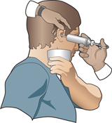
SC 6. The auditory canal ends at the tympanum. Can you hypothesize what the function of the tympanic membrane might be?
Self-Check Answers
SC 1. The pinna and auditory canal collect and direct sound waves.
SC 2. Cupping the ear helps to collect and direct more sound waves.
SC 3. The wax moistens the passage and eardrum for flexibility and collects debris.
SC 4. The auditory canal also channels out debris and directs sound to the tympanic membrane.
SC 5. The syringe is used to fill the auditory canal with warm water to float out pieces of wax and remove wax buildup.
SC 6. Sound waves cause the tympanum to vibrate, converting sound energy to mechanical energy.
To review the structures found in the outer ear and their functions, summarize your understanding by creating a table similar to the one below. Save the table in your course folder for later access.
THE STRUCTURES AND FUNCTIONS OF THE OUTER EAR
Structure |
Function(s) |
|
|
|
|
|
|
1.36. Page 3
Module 1—The Nervous System
The Middle Ear
oval window: a membrane-covered opening located between the chamber of the middle ear and the wall of the inner ear
The stapes (stirrup) is attached to it and transmits sound waves by it to the inner ear.
amplify: to increase the amplitude or the extent of a vibration; see intensity
ossicles: three tiny bones located in the middle ear that are connected to each other, to the tympanum, and to the oval window; involved in amplifying sound waves
malleus (hammer): the first in the sequence of the three ossicles located in the middle ear; connected to the tympanum and the second ossicle (incus); part of a lever system that amplifies sound waves
incus (anvil): the middle in the sequence of the three ossicles located in the middle ear; connected to the malleus and the third ossicle (stapes); part of a lever system that amplifies sound waves
stapes (stirrup): the third in the sequence of the three ossicles located in the middle ear; connected to the second ossicle (incus) and the oval window; part of a lever system that amplifies sound waves and causes vibrations in the fluid of the inner ear
The middle ear extends from the tympanum to the oval window. The structures of the middle ear are specialized to conduct and amplify the mechanical vibrations produced by sound waves. The tympanic membrane is connected to the ossicles: the malleus (hammer), incus (anvil), and stapes (stirrup).
When you have a cold and are suffering from a throat infection, your throat infection can quickly develop into a terrible earache. It may feel like your eardrum will break or burst from the pressure that is building in the middle ear. In some serious cases, ear infections can actually cause the eardrum to rupture. The rupturing and damage to the tympanic membrane can result in partial hearing loss.
The normal vibrations of the tympanic membrane cause the ossicles to act as a lever conducting and amplifying vibrations through the middle ear.
Do you remember what structures make up the middle ear? Be careful here. The diagram seems to show that the semicircularcanals are part of the middle ear. This is not so! The middle ear is bordered by the tympanum and the oval window.

Inquiry into Biology (Whitby, ON: McGraw-Hill Ryerson, 2007), 420, fig. 12.20. Reproduced by permission.
Eustachian tube: a tiny passageway extending from the middle ear to the throat (pharynx); a passageway through which viruses and bacteria can travel from the nose and mouth, resulting in an inner ear infection
The Eustachian tube plays an important role in equalizing air pressure on both sides of the tympanum.
Did you notice the Eustachian tube? Taking into account the location of the Eustachian tube, can you hypothesize what events may occur as a result of a throat infection to lead to the bursting of the eardrum? When you feel your eardrum “popping,” it is an indication that the pressures on each side of the eardrum are equalizing. This is common when flying in an unpressurized plane, swimming or diving under water, or travelling in the mountains. Yawning, chewing, or swallowing can help equalize the pressures.
At this time you may wish to make summary notes, a concept organizer, or a table of structures and functions like the one below. You may also wish to prepare a labelled diagram of the ear that shows all structures and functions. Save your work in the course folder.
THE STRUCTURES AND FUNCTIONS OF THE MIDDLE EAR
Structure |
Function(s) |
ossicles (malleus, incus, stapes) |
|
oval window |
|
Eustachian tube |
|
 Self-Check
To help you understand and apply the concepts on the outer and middle ear, complete the following questions. Remember to write complete explanations in sentence form and to use correct biological vocabulary. Check your answers and file your work in your course folder. If you are unsure about an answer, consult with your teacher.
SC 7. Describe what would happen if a person poked a hole in his or her tympanum while trying to remove ear wax.
SC 8. On a plane ride where the pilot is expecting to encounter significant turbulence, the flight attendants may give hard candies to the passengers. Explain the physiological basis of this.
SC 9. Propose a hypothesis to explain why humans do not have enormous, moveable pinnae like some animals do.
SC 10. Explain why middle ear infections are common.
Self-Check Answers
SC 7. If the tympanum was punctured, the sound waves entering the ear through the auditory canal would not effectively be transferred to the ossicles because the tympanum would not vibrate freely in the same way as it would if it were whole and intact. Also, the sound waves would not effectively be amplified by the tympanum. Note: Answering that the person would not be able to hear as well is true, but it is not an answer with sufficient detail for Biology 30.
SC 8. Hard candies are given to encourage swallowing. Swallowing keeps the Eustachian tubes open so that pressure on the inside of the eardrum can change to accommodate any changes on the outside of the eardrum due to the turbulence.
SC 9. Humans do not have big, moveable pinnae because they depend on other senses, such as sight, to a greater extent than they do on hearing.
OR
Humans have large cerebral lobes and have learned that they can channel the sound by moving their head and “cupping” the ear.
SC 10. Middle ear infections are common because microorganisms, such as bacteria and viruses, can reach the middle ear by entering at the nose and at the mouth, eventually making their way up the Eustachian tube by way of the throat.
1.37. Page 4
Module 1—The Nervous System
The Inner Ear
cochlea: contains the organ of Corti, which functions to convert the mechanical energy of sound waves into a nerve impulse
round window: a membrane-covered opening between the chamber of the middle ear and the inner ear located below the oval window
The round window functions in dissipating sound waves in the inner ear.
auditory nerve: a nerve composed of sensory fibres from the organ of Corti, the vestibular apparatus that conducts impulses to the temporal lobe of the cerebrum
Sound has been collected, transmitted, and amplified through the outer and middle ear, but you haven’t heard anything yet. The inner ear is responsible for converting mechanical stimulation to a nerve impulse to be communicated to the brain. The inner ear also converts mechanical stimulation into information about balance to be interpreted by the brain.
Can you remember which structures make up the inner ear? The inner ear is composed of two organs: one for hearing and one for balance. Note the structures for hearing, which include the oval window, cochlea, round window, and auditory nerve. Note also the utricle, saccule, and semi-circular canals, which all have functions in maintaining balance.
You may wish to record the information in summary notes, a concept map, or as additions to your diagram or table. Store your work in your course folder.
THE STRUCTURES AND FUNCTIONS OF THE INNER EAR
Structure |
Function(s) |
oval window |
|
cochlea |
|
round window |
|
auditory nerve |
|
Organ of Corti
Special parts of the inner ear must be functional in order to facilitate hearing. So far, you have not encountered the mechanoreceptors of these parts and how they function in hearing. Mechanoreceptors are specialized receptor cells that detect a stimulus, such as pressure or vibration.
As you study the parts of the inner ear, you will discover why people can lose their range of hearing as they age.
 Read
Read
Read the section about the inner ear and the organ of Corti on pages 420 and 421 of your textbook. Study “Figure 12.21” on page 420, which shows a cross-section of the cochlea and the location of the organ of Corti (blown up on the far left of the diagram). The receptors for sound waves are located inside the organ of Corti. Make summary notes or summarize the functions of the structures of the organ of Corti in a table similar to the following. Save your work in your course folder.
THE STRUCTURES AND FUNCTIONS OF THE ORGAN OF CORTI
Structure |
Function(s) |
basilar membrane |
|
hair cells |
|
tectorial membrane |
|
auditory nerve |
|
sensory neurons |
|
basilar membrane: one of two parallel membranes that comprise the organ of Corti in the inner ear
Hair cells are attached to the basilar membrane.
hair cells: sensory mechanoreceptors attached to the basilar membrane in the organ of Corti
tectorial membrane: one of two parallel membranes found in the organ of Corti
During the transmission of sound waves, the basilar membrane vibrates and causes the sensory hairs to flex against the tectorial membrane.
sensory neurons: nerve cells that are stimulated by hair cells in the organ of Corti to conduct messages toward the temporal lobe of the brain
The vibrating ossicles cause the oval window to vibrate. This causes pressure waves in the fluid-filled canals of the cochlea. The waves travel to the round window, which physically moves to equalize pressure. It is important to note how the structures in the cochlea function. The organ of Corti, resting on the basilar membrane, consists of hair cells, a tectorial membrane, and sensory nerve fibres. Imagine water plants moving with the current in a stream. Movement of the fluid in the inner ear causes vibration of the basilar membrane, which flexes the hair cells in the organ of Corti. The hair cells bend, like the plants in the stream, against the tectorial membrane. This mechanical action stimulates the dendrites of the sensory neurons and an electrochemical nerve impulse is initiated. The auditory nerve then transmits the impulse to the temporal lobe of the cerebrum where it will be interpreted as sound.
 Watch and Listen
To see this in action, visit the McGraw-Hill Ryerson online learning centre at www.albertabiology.ca. Navigate to the Student Resources, and select “Chapter 12” from the left column. Then choose “Animations.” Select “Effect of Sound Waves on Cochlear Structures.”
1.38. Page 5
Module 1—The Nervous System
 Try This
TR 1. To ensure that you understand the mechanisms of the inner ear involved in hearing, complete this drag-and-drop activity about the organ of Corti. If you have any questions about the inner ear, ask your teacher.
 Self-Check
The following Self-Check questions will help you review the concepts of the outer, middle, and inner ear. Consult with your teacher if you are not able to give complete answers. Store your answers in your course folder.
SC 11. Imagine that you are a sound wave. In order, describe the structures of the ear that you would encounter, starting with the outer ear and finishing with the organ of Corti.
SC 12. What happens to the energy of sound waves, which travel through air, once they reach the tympanum?
SC 13. How is the surrounding medium of the inner ear different from that of the middle ear? What needs to happen to the vibrations in the middle ear in order to accommodate this difference?
SC 14. What is the role of the hair cells located in the organ of Corti?
SC 15. Nerve impulses initiated in the organ of Corti are sent to which part of the brain?
Self-Check Answers
SC 11. In correct sequence, sound waves encounter the following structures:
pinna, auditory canal, tympanum, ossicles (malleus, incus, and stapes), oval window, cochlea, organ of Corti
SC 12. When the energy of sound waves comes in contact with the tympanum, it causes it to vibrate. Its vibrations are passed on and amplified by the neighbouring ossicles. At this point, the sound energy has been transformed into mechanical energy. Each bone vibrates more than the previous one so that the vibrations are amplified as they pass from one to the other. The last bone concentrates the vibrations onto the oval window. The vibrations, in turn, cause the fluid in the cochlea to vibrate, creating pressure waves that initiate an electrical nerve impulse in the auditory nerve.
SC 13. The middle ear is filled with air, whereas the inner ear is filled with fluid. In order to convert the sound vibrations of the middle ear into the pressure waves of the inner ear, the sound vibrations need to be amplified a great deal. It is much harder to move fluid than it is to move air.
SC 14. The role or function of the hair cells is to initiate a nerve impulse in the auditory nerve when they are stimulated by the pressure waves in the fluid of the inner ear.
SC 15. The auditory nerve transmits nerve impulses initiated by the hair cells to the temporal lobe of the cerebrum. Note: Biology 30 students require a more sophisticated and detailed answer than just simply “the cerebrum”!
 Watch and Listen
To consolidate your understanding of the concepts up to this point, go to the McGraw-Hill Ryerson online learning centre at www.albertabiology.ca. This site has many resources. Click on “Student Resources” in the column on the left side of the screen. Click on the “continue” button. You should now be on the main Inquiry into Biology page. Listed on the left side of the screen are many resources that are available to you. Click on “Essential Study Partner” in the left column. Then, click on “Essential Study Partner” in the white part of the screen. Next, click on the “Animals” icon. Choose “Sense Organs” from the list at the left, and then click on “6.81 Sounds Waves.” Watch an animated clip that shows how sound waves are captured, transformed into mechanical vibrations, amplified, and finally converted into nerve impulses that are transmitted to the brain—all by the different structures of the ear.
OR
You may want to watch a video about the anatomy and physiology of the ear. Watch the following segments of the video “The Ear: Hearing and Balance: 'Sound Sense'.”
- “Processing of Sound Waves”
- “Ear Structure”
- “Detailed Anatomy of the Ear”
- “Bio Fact: Bones of the Middle Ear”
- “Ear Function”
- “Detailed Physiology of the Ear”
Self-Check

Inquiry into Biology (Whitby, ON: McGraw-Hill Ryerson, 2007), 420, fig. 12.20. Reproduced by permission.
SC 16. Which three parts of the ear are involved in amplifying sound waves?
SC 17. The initiation of nerve impulses occurs in which structure?
SC 18. Which structure first directs sound waves into the ear?
SC 19. Which structure aids in the equalization of pressure between the outer and inner ear?
SC 20. Which structure is most affected if pressure is not equalized?
SC 21. Where is the organ of Corti located?
SC 22. Middle ear infections affect which structure?
SC 23. In the ear, which structure is considered analogical to the optic nerve in the eye?
SC 24. Where are the wax glands located?
SC 25. Which structure causes the ossicles to vibrate?
Self-Check Answers
SC 16. The auditory canal, tympanum, and ossicles amplify sound waves.
SC 17. The initiation of nerve impulses occurs in the cochlea; more specifically, the organ of Corti.
SC 18. The pinna directs sound waves into the ear.
SC 19. The Eustachian tube assists in balancing pressures between the outer and inner ear.
SC 20. The tympanum is most affected if the pressures are not equalized.
SC 21. The organ of Corti is located in the cochlea.
SC 22. Middle ear infections affect the tympanum.
SC 23. The auditory nerve is analogical to the optic nerve.
SC 24. The wax glands are located in the auditory canal.
SC 25. The tympanum causes the ossicles to vibrate.
1.39. Page 6
Module 1—The Nervous System
Hearing Pitch and Loudness
 Read
Read

© Eric Bechtold/iStockphoto

© Angel Herrero de Frutos/iStockphoto
pitch: corresponds to the frequency of a sound wave
frequency: number of wavelengths per given time; number of waves that pass a given point in a given time
loudness: subjective interpretation of sound intensity
You have likely been to a concert or listened to a MP3 player, or perhaps you play a musical instrument. If so, you are familiar with high-pitch (high frequency) and low-pitch (low frequency) sounds. However, do you know how or why you are able to hear high- or low-pitch sounds? How are you able to distinguish between yelling and whispering, or loudness?
To understand how people hear, read from “Frequencies of Sound” on page 421 through “The Perception of Sound” on page 424 in the textbook. When you have finished, make summary notes, a concept map, or a flow chart for your course folder.
 Try This
To assure your understanding of the concepts of pitch and loudness, answer the following questions in full sentences. If you are unsure of any of the concepts, consult with your teacher. File your exercise in your course folder.
TR 2. Define frequency.
TR 3. What is pitch? What is the relationship between pitch and frequency?
TR 4. What is the amplitude of a sound wave? How is the intensity or loudness of a sound related to the amplitude of a sound wave?
TR 5. Each of the sounds described below predominantly illustrates either pitch or loudness. Classify each as one or the other according to its main feature.
- the whine of a mosquito
- the sound of a low note on the piano
- the sound of a jackhammer breaking up a cement sidewalk
- the sound of jet plane taking off
- the growl of a dog
Loudness, Pitch, and the Organ of Corti
As you have read, the hair cells in the organ of Corti are sensitive to the frequency (pitch) as well as to the amplitude (loudness) of sound waves. The basilar membrane closest to the oval window (where frequency is 20 000) is narrow and stiff, as shown in the diagram below. It responds to high-frequency sound waves. The basilar membrane, by the apex of the cochlea (where frequency is 20), is wide and flexible. It responds to low-frequency sound waves.

As part of your teacher-marked assignment, you will complete a lab examining the differences between pitch and loudness. In preparation for this lab, notice in the diagram that the wider basilar membrane furthest from the oval window and closest to the tip of the cochlea responds to lower-frequency sound waves. Different hair cells are stimulated by different frequencies. In the diagram, the numbers correspond to the range of frequencies that humans can hear. In comparison to humans, dogs can hear very high sounds in the range of 40 000 Hz. Reflect on how this knowledge is used in the use of a dog whistle. Mice can hear in the range of 80 000 Hz. And elephants can hear sounds as low as 16 Hz. Hair cells at either end of the basilar membrane can be damaged over the years, and they do not regenerate. How would this damage affect your range of hearing as you age? If you guessed that the ability to hear high and low sounds deteriorates, you were right.
Try This

© Four Oaks/shutterstock
Choose one of the following Try This activities.
TR 6. Demonstrate your understanding of how sound is perceived by answering the following questions in complete sentences.
-
How is the brain able to perceive sounds of higher or lower pitch and softer or louder sounds?
-
Mice and dogs can hear sounds in the range of 40 000 to 80 000 Hz, whereas elephants hear sounds as low as 16 Hz. Describe how the cochlea of these animals might be modified in comparison to the human cochlea.
-
Why are elderly people often not able to hear very high-pitched sounds?
TR 7. Do questions 6, 18, and 19 on page 432 of the textbook.
1.40. Page 7
Module 1—The Nervous System
 Module 1: Lesson 6 Assignment
 Lab—Hearing: Pitch and Loudness
To apply your understanding of pitch and volume, open the Module 1: Lesson 6 Assignment that you saved to your computer earlier in this lesson. You will complete Part 1—Lab using the Frequency and Volume Gizmo. Read through the entire lab before beginning.
Have you ever blown a dog whistle? Could you hear it? Probably not, because the sound frequencies produced by a dog whistle are higher than the range of sounds humans can hear. You’ve probably observed animals that seemed to be agitated by sounds that you couldn’t hear. You may have heard an older person say “speak into my good ear.” The range of sounds that can be heard gets smaller as people age, especially if they are repeatedly exposed to loud sounds from lawnmowers, snowmobiles, heavy traffic, sirens, loud concerts, MP3 players, and televisions.
In this exercise you will test your hearing range by listening to low-, medium-, and high-frequency sounds. You will compare the relative loudness of sounds at each frequency to create an equal-loudness curve and a curve for your threshold of audibility.
Note: It is important for you to read the procedure for the lab beforehand so that you can choose your subjects and so that you can make an honest prediction as to how each of your subject’s results will compare with your own readings in terms of the sounds participants can detect.
Problem (Purpose)
What is your threshold of audibility for different frequencies, and how do your results compare to others?
Materials
No additional material are required other than access to the “Hearing: Frequency and Volume Gizmo.” Read the rest of the lab information before opening the gizmo.
You may be required to enter a username and password to access the gizmo. Contact your teacher for this information.
Prediction
If you are posting your equal loudness curve and your threshold of audibility curve, predict whether your classmates’ curves will be higher, lower, or the same as yours. Using what you have learned about the ear and hearing, explain why you think the curves will be as you have predicted.
OR
If you are using members of your family who are of different ages, predict whether their equal loudness and threshold of audibility curves will be higher, lower, or the same as yours. Using what you have learned about the ear and hearing, explain why you think the curves will be as you have predicted.
Procedure
Step 1: First, eliminate as much of the background noise where you are as possible. Background noise is the sounds coming from sources other than your computer’s speakers or headphones, such as a radio or a fan. Use headphones if you have access to them.
Be sure the system volume slider is set to 1 and click the “test” button. If the sound is too loud, turn down your system’s speakers or the computer’s volume and click “test” again. If the sound is too soft, turn up your speakers or adjust the system volume slider until a comfortable volume is reached.
Step 2: The pitch of a sound is related to its frequency, which is measured in hertz (Hz), or cycles per second.
Locate the 500-Hz slider, and move it to minus 20 decibels (dB). Click the “play” button for the 500-Hz frequency and listen. Did you hear anything? If so, was the sound loud or soft?
The decibel value for a sound wave is a ratio that is used to describe the loudness of that sound compared to a reference value. In this gizmo, a zero decibel sound has the same loudness as the reference sound. The more negative the decibel value, the softer the sound. Therefore, a negative 30 decibel sound is softer than a negative 20 decibel sound.
Step 3: Locate the slider for the 4000-Hz sound. (Note that this slider is between the slider for 1000 Hz and 5000 Hz.) Move this slider so that it has the same decibel value as the 500-Hz frequency. Play the 500-Hz sound, and then play the 4000-Hz sound. Does one tone seem louder than the other?
Step 4: How loud a sound actually seems, or the perceived loudness, depends on the way your ears function. Human ears can detect some frequencies better than others.
Adjust the 4000-Hz slider until it seems to have the same perceived loudness as the 500-Hz sound.
Step 5: Repeat Steps 3 and 4 for the remaining frequencies. Start the slider for each frequency at the same intensity as the 500-Hz slider, and then compare the sound to the 500-Hz tone. Move the slider for each frequency up or down as necessary until it has the same perceived loudness as the 500-Hz sound. Note that you may have difficulty hearing any sound at all for a few of the highest and lowest frequencies. This is most likely not because of a hearing problem, but because of the quality of your computer’s speakers.
Step 6: Press the “play full sequence” button, and listen to all the tones. Did each sound seem equally loud? If one sound seemed louder or softer than the others, adjust the slider for that frequency and press “play full sequence” again.
Step 7: Look at the positions of the sliders from left to right. Imagine connecting the sliders with a smooth curve. What shape would the curve have? This curve is called an equal loudness curve. Click “copy screen” to take a screen shot, and paste the image into a blank document. Post your graph on the discussion board and compare your curve to that of three or four classmates.
If it is not possible to compare your graph to classmates' graphs, help members of your family complete the procedure and make equal loudness curves. If possible, have a middle-aged person or a senior, as well as someone your own age and someone several years younger, participate in your lab. Paste all of the curves into your blank document so that you can submit these as data for your lab.
- Were all the curves similar?
- Did some people hear some frequencies differently from others?
Step 8: Now you will create an equal loudness curve for the faintest sound that you can hear at each frequency. For good results in this part of the experiment, the room should be as quiet as possible. By moving the 500-Hz slider and using the 500-Hz “play” button, experiment until you have found the lowest volume at which you can detect a 500-Hz tone. This sound intensity is your threshold of audibility for a frequency of 500 Hz.
Step 9: Predict what the intensity of a sound at your threshold of audibility for 4000 Hz will be. Using the same method as you did in Step 8, find the threshold of audibility for a 4000-Hz sound. Was your prediction correct, or was the intensity higher or lower than you expected?
Step 10: Find your threshold of audibility for each of the remaining frequencies. The equal loudness curve that results shows the lowest sound intensity that you can hear for each frequency in these conditions.
Step 11: Click “copy screen” and paste the screen shot into your document. Post this graph on the discussion board and compare your curve to that of three or four classmates. As before, if that is not possible repeat the exercise with the same people that you had participate in the activity for the equal loudness curve. How do their curves compare to yours? Note: When establishing the threshold of audibility curves, use the same computer and/or the same system settings to ensure a controlled experiment.
Observations and Data Collection
The properly identified and downloaded curves for you and your experimental subjects will make up your data.
Analysis and Discussion
What does your personal data indicate?
- Does your personal data, including both the equal loudness curve and the threshold of audibility curve, appear as a straight line? Why or why not?
- If it is not a straight line, which frequencies were easier to hear and which were more difficult to hear?
- Were there any frequencies that you could not hear at all?
- Compare the two curves of each of your subjects with your own curves. Are there any differences? If so, what does this tell you about their hearing?
- What parts of the ear are involved in the process of hearing the different pitches and varying levels of loudness?
- Compare the actual results to the predictions that you made before starting the experiment. Were your predictions correct? What factors might have made their curves different? Answer these questions in a paragraph form.
Conclusions
From your observations, what can you conclude from this experiment? In this section you should be able to answer the question that was posed in the Problem or Purpose section of this lab.
Copy all of your findings and answers for this lab into the space provided in the Module 1: Lesson 6 Assignment.
 Self-Check
To further check your understanding of the pitch and loudness, complete the five questions in the gizmo, and then check your answers.
When you have completed the lab, save your assignment in your course folder. You will receive instructions about when to submit your assignment to your teacher later in this lesson.
 Going Beyond
If you found the lab interesting, and you want to learn more about hearing loss, you may want to investigate tinnitus, which is a disorder experienced by many hearing impaired people.
1.41. Page 8
Module 1—The Nervous System
Homeostatic Imbalance in Hearing

© 2008 Jupiterimages Corporation
Healthcare professionals are very concerned that the present generation of young people will experience increased hearing loss as it ages. Deafness can be defined as one of two main types: conduction deafness or perception deafness.
deafness: any type of hearing loss, whether it is the inability to detect certain frequencies, the inability to detect very soft sounds, or the inability to detect any sound at all
conduction deafness: loss of hearing that results when the structures of the ear required to transmit and amplify vibrations (tympanum, ossicles, and oval window) are unable to do so; involves a malfunction in the conduction apparatus
perception (nerve) deafness: loss of hearing that results when the structures of the organ of Corti are unable to initiate nerve impulses
 Read
Read
From your reading of pages 423 and 424 and “Table 12.3,” make summary notes or a chart about the types and causes of deafness.
 Self-Check
SC 26. What is meant by the term deaf?
SC 27. State the parts of the ear that have been damaged or are non-functional in someone with
- perception (nerve) deafness
- conduction deafness
Self-Check Answers
SC 26. The term deaf refers to any type of hearing loss.
SC 27.
- In perception (nerve) deafness, any of the structures of the organ of Corti have been damaged, including the hair cells, the sensory neurons, the basilar membrane, the tectorial membrane, or the auditory nerve.
- In conduction deafness, structures such as the tympanum, any of the ossicles, or the connection between the stapes and the oval window may be damaged. In addition, things such as a buildup of wax in the auditory canal and against the tympanum can impede the conduction and amplification of sound in the inner ear.
Maintaining Balance
semicircular canals: three tubes that are situated at right angles to one another and contain mechanoreceptors that detect head and body rotation; responsible for dynamic balance or rotational equilibrium (rotational equilibrium dynamic balance), which is the balance that is established in response to the head and body being moved or rotated
utricle and saccule: tiny chambers in the inner ear that contain otoliths and hair cells, which respond to changes in head position with respect to gravity and movement in one direction; responsible for static balance or gravitational equilibrium
dynamic balance (rotational equilibrium): balance resulting when the head and body are moved or rotated
static balance (gravitational equilibrium): balance resulting from changes in the position or the movement of the head in one direction; usually in response to gravity
otolith: a tiny particle of calcium carbonate found in the utricle and saccule that contacts the hair cells of these structures and stimulates them
vestibule: the fluid-filled area of the inner ear located between the semicircular canals and the cochlea
The vestibule contains the utricle and saccule.
The ear has an important function in your ability to maintain balance. The inner ear has three additional structures known as the semicircular canals, the utricle, and the saccule that are responsible for maintaining balance.
When you twirl on the dance floor, spin in a rotating ride at the midway, or suffer through a rough boat ride, the semicircular canals are working overtime. This is called dynamic equilibrium or rotational equilibrium.
That queasy feeling you experienced when you pulled ahead of the truck slipping backwards on the icy hill and that feeling you get when a fast-moving elevator comes to an abrupt stop are the result of nerve impulses from the utricle and saccule. This is called static equilibrium or gravitational equilibrium. The utricle and saccule have otoliths, small granules of calcium carbonate that are pulled against the hair cells in the utricle and saccule when you move your head forwards or backwards. This mechanical stimulation starts a nerve impulse. Some impulses from the vestibule travel to the spinal cord, where body position can be adjusted by reflex action. Other impulses are sent to the cerebellum, where other reflexive muscular coordination occurs. Further impulses move to higher centres in the cerebrum involved in the control of eye movement. Input from the eyes is very important in maintaining balance. To show this, try closing your eyes and standing on one leg!
Read
At this point in your study of the ear, it is important to make sure that you include diagrams like the ones on pages 420 and 421 of your textbook. You may choose to combine the diagrams with a table listing the functions of each part that you have labelled. Save your work in your course folder.


left: © Robert J. Daveant/shutterstock; middle: © Melissa Schalke/shutterstock; right: © Carolina/shutterstock
To understand how the body maintains balance, choose one of the following two options. You may summarize the information in any format you choose and store this information in your course folder.
![]() Read pages 424 and 425 in the textbook.
Read pages 424 and 425 in the textbook.
OR
- “Balance”
- “Bio Review: Utricle and Saccule”
- “Dynamic Equilibrium”
- “Spins in Figure Skating”
Self-Check
Test your understanding of the concepts by completing the following questions. Save your work in your course folder. If you do not understand a particular concept, consult your teacher.
SC 28. Describe the structures of the inner ear that are involved in maintaining balance.
SC 29. Explain how the structures of the inner ear allow for dynamic (rotational) equilibrium.
SC 30. Explain how static (gravitational) equilibrium is attained.
Self-Check Answers
SC 28. There are three structures of the inner ear involved in maintaining balance. These structures include the three fluid-filled semicircular canals situated at right angles to each other and, just below them, the fluid-filled utricle and saccule, which contain tiny rock-like granules of calcium carbonate called otoliths.
SC 29. The balance established while moving the head forward and backward is called dynamic (rotational) equilibrium. At the base of each of the semicircular canals is a jelly-like covering called a cupula. The tiny hairs of the hair cells in the semicircular canals, better known as stereocilia, stick into the cupula. When the body rotates, the fluid inside the semicircular canals moves and bends the stereocilia. The hair cells receive this information and send it to the brain.
SC 30. A person who is lying down or not moving is said to be in a static position. The movement, or lack of movement, of the fluid in the inner ear tells the brain whether a person is standing, lying down, hanging upside down, etc. When an individual is moving around, calcium carbonate crystals (otoliths) in the utricle and saccule shift position and contact the stereocilia of these structures. This contact results in nerve impulses being transmitted to the brain, indicating that the individual is changing positions. When the person is in an established position, the crystals stop rolling around. The sensory neurons in the vestibule of the utricle and saccule do not send any impulses to the brain, thereby indicating that the individual is in a static position once again.
 Try This
You will need somebody to help you with this exercise. A friend or a member of your family will do!
Step 1: Have your partner sit in a swivel chair with his or her head held in an upright position. Get your partner to raise his or her legs up off of the ground. Rotate the chair in a clockwise direction, but not too quickly. After 20 rotations, stop the chair and get your partner to stand. Be prepared to support or catch your partner! Make observations.
Step 2: Allow your partner to recover for about 5 minutes. Repeat the steps, but, this time, rotate the chair in a counterclockwise direction. Make sure your partner is holding his or her head straight. What did you observe this time?
Step 3: Allow your partner to recover for about 5 minutes.
Step 4: Have your partner sit in the swivel chair, but this time with his or her head tilted to the right. Rotate the chair clockwise 20 times. Ask your partner to stand and hold his or her head upright. What happens?
TR 8. What kind of evidence did you collect in your experiment that would suggest that the fluid in the semicircular canals continues to move even after you have stopped rotating the chair?
TR 9. Describe how the semicircular canals detect changes in motion during a rollercoaster ride.
TR 10. Are the utricle and saccule involved in maintaining balance during a rollercoaster ride?
1.42. Page 9
Module 1—The Nervous System
 Self-Check
This Self-Check reflects the kind of questions that you will encounter on the Diploma Exam. The questions include both numerical-response and multiple-choice questions. Discuss any concerns with your teacher. This is a good opportunity to assess your understanding of the structures and functions of the ear.
Use the following diagram to answer the next two questions.

Inquiry into Biology (Whitby, ON: McGraw-Hill Ryerson, 2007), 420, fig. 12.20. Reproduced by permission.
SC 31. Match the following structures with those labelled on the picture of the human ear. Record the number from the diagram in the space that corresponds below. (Record all four digits of your answer in the spaces below.)
semicircular canals
tympanum
middle ear
cochlea
SC 32. The structure that is primarily responsible for maintaining dynamic equilibrium is represented by
- 1
- 2
- 3
- 4
SC 33. Which pair gives an INCORRECT function for the structure(s)
- Eustachian tube – equalization of pressure
- cochlea – initiation of nerve impulses
- round window – dissipation of pressure waves
- utricle and saccule – dynamic equilibrium
SC 34. Place the following events in the correct sequence after the first event stated below. Record the four digits of your answer below.
First Event: Sound waves push against the tympanum.
- Tiny hair-like cells in the organ of Corti respond to vibrations by stimulating sensory fibres.
- Vibrations are amplified by the ossicles.
- The basilar membrane begins to vibrate.
- The oval window receives vibrations from the ossicles
________________________________________
Use the following information to answer the next two questions.
| A cochlear implant is very different from a hearing aid. Hearing aids amplify sound. A cochlear implant is designed to compensate for damaged or non-functional parts of the inner ear. When hearing is functioning normally, complex parts of the inner ear convert sound waves in the air into nerve impulses. These impulses are then sent to the brain, where they are interpreted and recognized as sound. A cochlear implant works in a similar manner. It electronically finds useful sounds and then sends impulses to the brain. Sounds heard through an implant may sound different than sounds heard without, but it enables many people to communicate orally in person and over the phone who would not be able to do so without such a device. |
SC 35. Identify the specific structure(s) of the inner ear that the cochlear implant is intended to replace.
- tympanic membrane
- hair cells located on the basilar membrane
- round window
- ossicles
SC 36. Identify the specific structures of the ear that have the same function as a hearing aid.
- tympanic membrane and ossicles
- hair cells and sensory fibres on the basilar membrane
- round and oval windows
- vestibular apparatus
SC 37. Which of the following statements is INCORRECT with respect to hearing in humans?
- The hair cells of the organ of Corti are able to distinguish both the frequency (pitch) and amplitude (loudness) of sound waves.
- The hair cells of the organ of Corti are able to detect sound within the frequency range of 0 Hz to 20 Hz.
- High-frequency sounds most strongly stimulate the hair cells closest to the oval window.
- Low-frequency sounds most strongly stimulate the hair cells farthest from the oval window.
Self-Check Answers
SC 31. 2, 4, 1, 3 (in that order only)
SC 32. 2 (semicircular canals)
SC 33. D
SC 34. 2, 4, 3, 1 (in that order only)
SC 35. B
SC 36. A
SC 37. B
1.43. Page 10
Module 1—The Nervous System
 Reflect and Connect
This module introduced you to the nervous system and communication.
In the Big Picture at the beginning of this module, you experienced various forms of communication, both external and internal. Hearing is a very important part of communication. If perception of sound had not been communicated to your brain, how would you have interpreted the events that occurred while you were in the room with your friends?
 Module 1: Lesson 6 Assignment
In this part of your assignment, you will respond to the issue of noise-induced hearing loss. This work will involve an open-response essay like on the Diploma Exam.
Retrieve the copy of the Module 1: Lesson 6 Assignment that you saved to your computer earlier in this lesson. Complete Part 2—Closed Written Response. Save your completed assignment in your course folder. You will receive instructions about when to submit your assignment to your teacher later in this lesson.
Reflect on the Big Picture

©olly/shutterstock
If you were not able to hear laughter, people talking, and music playing, what might your perceptions have been like in the Big Picture at the beginning of this module? How does music at the beginning of a movie help to communicate the tone of movie? What kinds of music communicate dangerous, scary, funny, or romantic scenes? Before a big game, athletes often use music for motivation. How do you think Beethoven, who was deaf, was able to successfully convey all of the moods in his music?
Sounds in today’s very noisy world contribute to hearing loss in many people. Knowing the things you have learned in this lesson, you may be more aware of the volume of the music at the next concert or wedding you attend. Reflect on the precautions that you could take to protect your ears and your hearing the next time you mow the lawn, ride a snowmobile, listen to your MP3 player, or use power machinery. What do you notice when you shut the machine off? Could this be an indication of what you should do to safeguard your hearing?
In the Get Focused section of this lesson, you could hear the mosquito ring tone of your phone. As you grow older, will you always be able to hear this ring tone, the music you love, the sounds that ensure your survival, or those that bring you pleasure? Judging by your own habits when it comes to hearing, do you think that you might have to wear a hearing aid at some point in your lifetime?
Module 1: Lesson 6 Assignment
In the last section of your assignment, you will research some technologies available to enhance hearing. You will consider how they work and what part or function of the ear they are designed to assist.
Retrieve the copy of the Module 1: Lesson 6 Assignment that you saved to your computer earlier in this lesson. Complete Part 3—Hearing Technologies Research. Save your completed assignment in your course folder. You will receive instructions about when to submit your assignment to your teacher later in this lesson.
 Discuss
Consider the following scenario:
Courtesy of Library Services for the Deaf & Hard of Hearing
| Ethan was very excited to have landed a job as an assistant baggage handler at the airport. He got to drive the baggage train and load and unload suitcases and parcels from airplanes. However, he found that the ear protection that he was supposed to wear was hot and cumbersome. He took it off whenever he could, and sometimes he forgot to put it back on when the big jets came in or took off. After two years on the job, he found that he was having difficulty hearing. It was suggested that he purchase a very expensive new type of hearing aid that would greatly improve his hearing. Ethan was very upset because his medical plan did not pay for hearing aids. |
Do you think hearing aids should be covered by medical plans? Keeping Ethan’s story in mind, suggest several reasons why they should and several reasons why they should not. Using the discussion board, share and discuss the reasons supporting your argument as well as those that counter your argument. Share your final justification with your teacher. Save a copy of your work in your course folder.
Module 1: Lesson 6 Assignment
Submit your completed Module 1: Lesson 6 Assignment to your teacher for assessment.
1.44. Page 11
Module 1—The Nervous System
 Lesson Summary
In this lesson, the following focusing questions were investigated:
-
What are the major parts of the ear that facilitate your response to sound in the environment?
-
How do the structures of the ear impact your ability to maintain balance within your changing environment?
You have explored the structures and functions of the outer, middle, and inner ear. Both the cochlea, adapted for hearing, and the vestibular apparatus, adapted for maintaining balance, rely on specialized hair cells to change the mechanical energy of sound or movement to the electrochemical energy of a nerve impulse. The structures of the outer and middle ear conduct and amplify sound waves so that pressure waves can be created in the cochlea of the inner ear. The frequency of a sound wave results in your perception of pitch, whereas the amplitude (height) of the sound wave results in your perception of loudness. Conduction deafness and perception (nerve) deafness result when there is a dysfunction in the hearing apparatus. Both dynamic and static balance are maintained by the vestibular apparatus, which includes the semicircular canals, the utricle, and the saccule. Technologies, such as hearing aids and cochlear implants, have been developed to help with hearing loss.
The questions on pages 432 and 433 of your textbook provide an excellent opportunity to review and practise your knowledge and skills dealing with sensory reception. You may wish to do a selection of these questions as review. Your teacher can provide feedback on your answers.
Continue your research about Alzheimer’s disease. Are there any connections between this disease and hearing loss or balance problems?
Lesson Glossary
Consult the glossary in the textbook for other definitions that you may need to complete your work.
amplify: to increase the amplitude or the extent of a vibration; see intensity
auditory canal: a short channel that funnels sound waves from outside the ear to the tympanum and amplifies sounds
auditory nerve: a nerve composed of sensory fibres from the organ of Corti, the vestibular apparatus that conducts impulses to the temporal lobe of the cerebrum
basilar membrane: one of two parallel membranes that comprise the organ of Corti in the inner ear
Hair cells are attached to the basilar membrane.
cochlea: contains the organ of Corti, which functions to convert the mechanical energy of sound waves into a nerve impulse
dynamic balance (rotational equilibrium): balance resulting when the head and body are moved or rotated
Eustachian tube: a tiny passageway extending from the middle ear to the throat (pharynx); a passageway through which viruses and bacteria can travel from the nose and mouth, resulting in an inner ear infection
The Eustachian tube plays an important role in equalizing air pressure on both sides of the tympanum.
frequency: number of wavelengths per given time; number of waves that pass a given point in a given time
hair cells: sensory mechanoreceptors attached to the basilar membrane in the organ of Corti
incus (anvil): the middle in the sequence of the three ossicles located in the middle ear; connected to the malleus and the third ossicle (stapes); part of a lever system that amplifies sound waves
inner ear: one of the three regions of the ear that is located deepest into the head and consists of a fluid-filled chamber containing the semicircular canals, the utricle and saccule, and the cochlea
intensity: measurement of the difference between compressed and rarefied areas of a sound wave used in physics; corresponds to amplitude of a wave
loudness: subjective interpretation of sound intensity
malleus (hammer): the first in the sequence of the three ossicles located in the middle ear; connected to the tympanum and the second ossicle (incus); part of a lever system that amplifies sound waves
middle ear: one of the three main regions of the ear, which begins just past the tympanum and consists of a chamber containing three tiny bones (ossicles) called the malleus (hammer), the incus (anvil), and the stapes (stirrup); leads into to a minute opening called the Eustachian tube
organ of Corti: a structure of the inner ear that contains hair cells that detect vibrations in the fluid of the inner ear and initiates a nerve impulse that is transmitted to the auditory nerve
ossicles: three tiny bones located in the middle ear that are connected to each other, to the tympanum, and to the oval window; involved in amplifying sound waves
otolith: a tiny particle of calcium carbonate found in the utricle and saccule that contacts the hair cells of these structures and stimulates them
outer ear: one of the three main regions of the ear, which consists of the pinna and the auditory canal and ends at the tympanum or eardrum
oval window: a membrane-covered opening located between the chamber of the middle ear and the wall of the inner ear
The stapes (stirrup) is attached to it and transmits sound waves by it to the inner ear.
pinna: the outer flap of the ear that is composed of skin and cartilage and shaped so that it enhances sound vibrations and funnels them into the auditory canal
pitch: corresponds to the frequency of a sound wave
round window: a membrane-covered opening between the chamber of the middle ear and the inner ear located below the oval window
The round window functions in dissipating sound waves in the inner ear.
semicircular canals: three tubes that are situated at right angles to one another and contain mechanoreceptors that detect head and body rotation; responsible for dynamic balance or rotational equilibrium (rotational equilibrium dynamic balance), which is the balance that is established in response to the head and body being moved or rotated
sensory neurons: nerve cells that are stimulated by hair cells in the organ of Corti to conduct messages toward the temporal lobe of the brain
sound: pressure disturbance beginning at a vibrating object (source) and spread out by a medium such as air
sound wave: a series of compressions and rarefactions resulting in an S-shaped curve or sine wave
stapes (stirrup): the third in the sequence of the three ossicles located in the middle ear; connected to the second ossicle (incus) and the oval window; part of a lever system that amplifies sound waves and causes vibrations in the fluid of the inner ear
static balance (gravitational equilibrium): balance resulting from changes in the position or the movement of the head in one direction; usually in response to gravity
tectorial membrane: one of two parallel membranes found in the organ of Corti
During the transmission of sound waves, the basilar membrane vibrates and causes the sensory hairs to flex against the tectorial membrane.
tympanum: a round elastic structure located in the middle ear that vibrates in response to sound waves; also called the eardrum or tympanic membrane
utricle and saccule: tiny chambers in the inner ear that contain otoliths and hair cells, which respond to changes in head position with respect to gravity and movement in one direction; responsible for static balance or gravitational equilibrium
vestibule: the fluid-filled area of the inner ear located between the semicircular canals and the cochlea
The vestibule contains the utricle and saccule.
vestigial: small or imperfectly developed; often referring to a part or organ that was once more fully developed in an earlier evolutionary stage, but is now non-functional
1.45. Lesson 7
Module 1—The Nervous System
Lesson 7—the Nerve Impulse—Transporting the Message
 Get Focused

© Ricardo Verde Costa/shutterstock
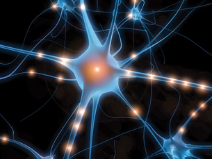
© Sebastian Kaulitzki/shutterstock
Your cellphone is ringing—how did you know? At this point in the unit, you should be aware that the retina in your eye and the cochlea in your ear are detecting the flashing light and the ringing noise.
You have learned that the sense organs are responsible for detecting changes in the environment and communicating this information to the brain. You now know that the neuron facilitates communication through sensory pathways that connect sensory receptors to the lobes of the brain. Interneurons and motor neurons also play a role in processing information and communicating information to the effectors.
In this lesson you will explore how this special type of cell, the neuron, which makes up the pathways and structures of the nervous system, communicates messages through its cell structure.
You will also examine how communication is interrupted by diseases, such as multiple sclerosis and Alzheimer’s disease. This lesson is a significant opportunity for you to explore the nerve impulse as a source of communication interruption in Alzheimer’s disease as you continue work on your Module Assessment.
In this lesson you will investigate the following focusing question:
- How does the structure of a neuron facilitate reception and transmission of a nerve impulse to the synaptic gap?
Lessons 7 and 8 present concepts that are both interesting and challenging. The suggested readings will help familiarize you with the lesson content and support your lesson and Module Assessment work.
 Module 1: Lesson 7 Assignment
Your teacher-marked Module 1: Lesson 7 Assignment requires that you complete and submit the following:
- Part 1—Action Potential OR Part 2—Action Potential
- Part 3—Reflect and Connect
Download a copy of the Module 1: Lesson 7 Assignment to your computer now. You will receive further instructions about how to complete this assignment and when to submit your work for assessment later in the lesson.
You must decide what to do with the questions that are not marked by the teacher.
Remember that these questions provide you with the practice and feedback that you need to successfully complete this course. You should respond to all the questions and place those answers in your course folder.
In addition to the lesson work that you store in your course folder, you will complete three types of questions in the Lesson 7 Assignment. In the first part of the assignment, you will analyze a graph of action potential and apply your understanding to a series of questions based on the principles illustrated in the graph. In the second part you will design a flow chart summarizing the events of the action potential. The third part of the assignment is a question similar to the closed-response written question on Diploma Exams. In this question you will apply your understanding of the principles that you have learned in this lesson to analyzing data given in the question. You will then answer a series of questions based on the data.
You will continue to add all lesson work and reflections to your course folder for later review. In addition to the assignments given in the lesson, there are several questions on page 384 and pages 402 and 403 in your textbook that you may choose to do to practise your skills when you have completed this lesson. These questions provide you with the practice and feedback that you need to successfully complete this lesson. Your teacher can give you feedback on your responses to these questions. More questions are available in The Key, the publication of Diploma Exam-style questions available from the Learning Resources Centre (LRC) or your local bookstore.
You will continue to work on your Module Assessment throughout this lesson. You will want to focus particularly on communication involving the brain and how transmission of the message in neurons of the brain can be interrupted.
1.46. Page 2
Module 1—The Nervous System
 Explore
The Neuron at Rest

left: © Laurin Rinder/shutterstock; right: © Bairachnyi Dmitry/shutterstock
In the previous lessons you examined how communication pathways, called sensory pathways, send information about taste, smell, touch, sight, and sound to the brain. Different lobes of your brain process this information, and motor pathways communicate appropriate responses to your effectors. In your mind, review how this works by adding information that you recall to the summary flow chart and relating it to the diagram below:
sensory receptors → sensory pathway → lobes of brain → motor pathways → effectors
Now you will examine some characteristics of the special type of cell, the neuron, which makes up the nerves in these pathways. As part of this exploration you will need to understand how messages are communicated through the neuron from dendrite to terminal end. To do this, you must investigate the characteristics of the neuron's membrane and explore the role of sodium and potassium ions as well as negatively charged particles, such as proteins and chloride ions, that surround the neural membrane.
 Read
Read
membrane potential: a form of potential energy resulting from the separation of charges between the inside and the outside of a cell membrane; voltage across the cell membrane
voltage: electrical potential difference across a membrane as measured by a voltmeter
polarized membrane: the state of the cell membrane in an unstimulated neuron in which the inside of the neuron is negatively charged in comparison to the outside of the neuron; the resting state of a membrane averaging approximately – 70 mV
polarization: the process of generating a resting membrane potential averaging approximately – 70 mV
To understand the characteristics of a neuron at rest and a nerve impulse, read "The Nerve Impulse" on pages 372 to 374 in the textbook. Study “Figure 11.12” on page 374 of your textbook, which describes how the sodium-potassium ion pump works. Summary notes and a chart similar to the one on page 374 will prove to be invaluable in mastering these concepts. Make sure you include the concepts of membrane potential, voltage, polarized membrane, polarization, depolarization, and repolarization in your work.
 Watch and Listen
To further explore the role of ions in establishing a resting or polarized state, go to the McGraw-Hill Ryerson online learning centre at www.albertabiology.ca. Navigate to Student Resources, choose “Chapter 11” from the menu on the left, and then select “Animations.” Choose the animation called “Sodium-Potassium Exchange Pump” from the list. This is a challenging and important concept. Be sure that you are able answer the following questions that are based on the animation:
- Identify two characteristics of active transport.
- In which direction are sodium ions moved across the neuron membrane by the sodium-potassium exchange pump?
- In which direction are the potassium ions moved?
- Where is the carrier protein of the ion exchange pump located?
- What causes the shape changes in the carrier protein?
- Where is ATP used?
resting membrane potential: the voltage that exists across a cell membrane during the resting state of an excitable cell, such as the neuron; averages around – 70 mV, but may range from – 50 to – 200 mV depending on the cell
You should now know that sodium ions become more concentrated outside the neuron and potassium ions become more concentrated inside the neuron. The net result is that the interior of the neuron (intracellular fluid) becomes negatively charged compared to the exterior (extracellular fluid), which becomes positively charged. When this occurs, the neuron is said to be polarized, or to have a polarized membrane. This can be verified by inserting a tiny electrode into the axon of the neuron and touching another to its surface. Usually there is a difference in charge or a resting membrane potential of approximately – 70 mV. This may vary from cell to cell and in different situations. Use the animated illustration of the polarized or resting neuron to review these concepts.
Where is the greatest concentration of the red circles, which represent the sodium ions? If you guessed outside the membrane, you were right. Where is the greatest concentration of the blue circles, which represent the potassium ions? If you guessed inside the membrane, you were correct. Besides movement of potassium ions by the ion exchange pump, what other ways can you see in the diagram that facilitate movement of potassium ions towards the outside of the cell? If you guessed diffusion and carrier molecules, you were correct. What do the black circles represent? If you guessed chloride ions, you were correct. Why are there no black circles on the exterior of the neuron? If you guessed that the membrane is impermeable to chloride ions, you were correct. What is the net charge on the exterior of the axon membrane? If you guessed positive, you were correct. What is the net charge on the interior of the axon? If you guessed negative, you were correct.
You may wish to review the concepts of diffusion, carrier molecules, permeability, and impermeability by using the Internet or reviewing your Science 10 materials.
Watch and Listen
The following segments of “Nerve Impulse Conduction: Dentists Calm Your Nerves” may help you to understand the processes of message transmission:
- “Bio Review: Neuron Structure and Function”
- “Ion Movement”
- “Ion Distribution”
- “Bio Simulation: Establishment of Membrane Potential”
 Self-Check
In your own words, answer the following questions in order to check your understanding of the concepts to this point. When you have finished, check your answers, make any corrections, and file your work in the course folder before you move on in the lesson.
SC 1. Define the term resting state as it is used to describe a neuron.
SC 2. Explain what the resting membrane potential is and why it is significant to the functioning of the neuron.
SC 3. Identify and explain the three factors that contribute to maintaining the resting membrane potential.
SC 4. Describe the distribution of sodium ions, potassium ions, and negatively charged particles in a resting neuron.
SC 5. What is meant by a “resting potential of – 70 mV”?
Self-Check Answers
SC 1. The neuron’s resting state is defined as the period when no nerve impulse is being generated.
SC 2. The charge difference across the neuron membrane in a non-stimulated, polarized, or resting neuron is called the resting membrane potential. The resting membrane potential is approximately – 70 mV, with the outside of the membrane having a positive net charge relative to the inside, which would have a negative net charge. The resting membrane potential is significant because it provides energy for the generation of a nerve impulse in response to an appropriate stimulus.
SC 3. Neurons generate a resting membrane potential (polarized state) by
- the selectively permeable membrane of the neuron being impermeable to the negatively charged particles, namely chloride ions and negatively charged proteins
- the sodium-potassium ion exchange pump, which uses energy to pump three sodium ions out of the neuron and two potassium ions into the neuron, resulting in an uneven distribution of positive charge inside and outside the membrane.
This buildup of positive charge on the outside creates an electric potential.
- special transit proteins that allow potassium ions to diffuse out of the neuron.
Fewer sodium ions are allowed to diffuse into the neuron, resulting in more positive charges outside the neuron than inside the neuron.
SC 4. In a resting neuron, there are more sodium ions on the outside of the neuron membrane than inside, and there are more potassium ions on the inside than there are on the outside of the neuron membrane. There are more negatively charged protein particles and chloride ions on the inside than there are on the outside of the neuron membrane. Therefore, the resting neuron has a net positive charge on the outside of the membrane, and a net negative charge on the inside of the neuron.
SC 5. A “resting potential of – 70 mV” means that the difference between the net positive charge on the outside and the net negative charge on the inside is – 70 mv.
1.47. Page 3
Module 1—The Nervous System
 Watch and Listen
Communication through the neuron involves a series of action potentials. To master the concepts of neuron communication, watch the following segments of “Nerve Impulse Conduction: Dentists Calms Your Nerves.” Your teacher can help you understand any of these concepts if you feel that you need further clarification after watching the segments.
- “Recording Impulse Conduction”
- “Bio Discovery: The Action Potential”
- “Bio Review: Depolarization”
The Action Potential
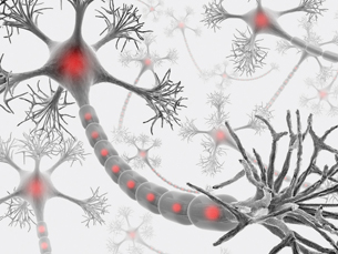
© iDesign/shutterstock
© Friax74/Fotolia
When a neuron becomes sufficiently stimulated by a threshold stimulus, the point of stimulation becomes depolarized and the depolarization spreads along the length of the unmyelinated neuron. This depolarization is created by a rapid change in membrane permeability and a corresponding change in the balance of ions maintained at the resting state. Depolarization can be likened to a burning fuse. The flame, like the wave of depolarization, progresses along the wick in one direction. However, in the fuse, the wick is burned and cannot be used again. In the neuron, after the wave of depolarization, an immediate recovery period called repolarization occurs.
action potential: the change in charge that occurs when the gates of the potassium ion channels close and the gates of the sodium ion channels open; a large depolarization event that is conducted along the membrane of a nerve cell or a muscle cell
threshold stimulus: weakest possible stimulus that is needed to initiate a nerve impulse
depolarization: the loss or reduction of the negative resting membrane potential
repolarization: restoring the resting membrane potential (– 70 mV) from the depolarized state
 Read
Read
To understand the next steps in communication, read “Action Potential” on pages 374 to 377 in your textbook. Making summary notes to be stored in your course folder will prove to be invaluable in mastering these concepts. You may wish to create your own illustration of these events and add it to your course folder for review.
Watch and Listen
You may wish to review the “Bio Discovery: The Action Potential” segment in the video “Nerve Impulse Conduction: Dentists Calms Your Nerves.”
For more review of the action potential, go to the McGraw-Hill Ryerson online learning centre at www.albertabiology.ca. Navigate to the Student Resources, and choose “Chapter 11” from the menu on the left side of the screen. From there, choose “Animations.” Select the animations called “Action Potential Propagation in an Unmyelinated Axon” and “Voltage Gated Channels and the Action Potential.” After watching the animations, answer the questions listed below.
Questions for “Action Potential Propagation in an Unmyelinated Axon”
- What is the net charge on the outside of a resting neuron?
- What is the net charge on the inside of a resting neuron?
- Describe the action potential.
- What keeps the action potential going along the axon of the neuron?
- What are localized electrical circuits?
- Why does the action potential proceed only in one direction?
- What is an absolute refractory period?
Questions for “Voltage Gated Channels and the Action Potential”
- Draw an illustration of the voltage-gated Na+ channel and the inactivation gate.
- Draw an illustration of the voltage-gated K+ gate.
- Describe how depolarization occurs and what happens in repolarization.
The Refractory Period and the All-or-None Principle
refractory period: the short time immediately after an action potential in which the neuron cannot respond to another stimulus; period of time it takes to re-establish the net positive charge on the outside of the neuron and the net negative charge on the inside of the neuron, where there are more sodium ions on the outside and more potassium ions on the inside of the neuron
threshold potential: the smallest change in the membrane potential of a cell membrane that is needed to initiate an action potential; approximately – 55 mV
all-or-none response: action that occurs either completely or not at all, such as the generation of an action potential by a neuron
Throughout this module, you have discovered that normal communication involves periods of interruption. In this case, while repolarization was occurring, there was no chance for communication. Another sodium inrush could not occur. This brief period when no stimulus can elicit a response is called the refractory period. Its duration is usually only 1 to 2 ms (milliseconds).
The stimulus that begins the influx of sodium ions and starts depolarization has to be of certain intensity in order to open the voltage-gated sodium channels. Once this voltage shift is reached, a wave of depolarization begins. It doesn’t matter if the voltage shift is higher, because once the gates are open, a wave of depolarization occurs. It is just like lighting the wick in the fuse analogy—either it ignites and begins to burn, or it does not ignite and the wick does not burn. A wave of depolarization is much the same. If the stimulus is not strong enough to open the gates in the neuron membrane, nothing happens. There isn’t a wave of depolarization—so there isn’t a nerve impulse. The voltage shift needed to open the sodium gates is called the threshold potential. This ability of the neuron to only respond to a disturbance in electrical charge of a specific threshold value is called the all-or-none response. Like the wick that either did or did not burn, the neuron either transmits the wave of depolarization or it doesn’t. In an all-or-none response, there are no in-betweens. To help illustrate this concept, consider another analogy—dominos that have been stood up in a line. Once you apply sufficient pressure to the first domino, all of the dominos will fall—there’s no stopping them. Once the threshold potential is reached, there is an all-or-none response that cannot be stopped, resulting in a wave of depolarization and impulse.
Watch and Listen
To view these events, watch the following segments of “Nerve Impulse Conduction: Dentists Calms Your Nerves.”
- “All-or-None Principle”
- “Nerve Impulse”
1.48. Page 4
Module 1—The Nervous System
 Self-Check
© Andriy Solovyov/shutterstock
SC 6. One way to model the action potential is to line up several dominoes and initiate a cascade event, in which each successive domino knocks down the next domino.
- In this model, the hand provides the initial energy. What provides the initial energy in a neural impulse?
- The finger has to contact the first domino just hard enough to get it to fall. Which action does this represent in a real neuron?
- Once the dominoes start to fall, they all fall in succession. What does this action represent in the real neuron?
- The dominoes always fall in one direction. Contrast this with the direction of impulse transmission in a real neuron.
- No matter how many times the dominos fall, they always move at the same speed and intensity. What principle does this represent in the real neuron?
Check your answers before proceeding further in this lesson.
Self-Check Answers
SC 6.
- To start an action potential or a nerve impulse, a stimulus such as light energy, sound vibration, hot or cold, or a chemical that is intense enough to cause the neuron to reach threshold potential (to cause enough sodium gates to open to begin depolarization) is needed.
- This demonstrates part of the all-or-none response—the action potential is either generated by the stimulus or it isn’t (just like the domino either falls or it doesn’t). The stimulus must reach threshold potential or depolarization will not occur.
- In a real neuron, this would be described as a wave of depolarization along an axon or the propagation of the action potential along the neuron. The impulse travels in only one direction in a neuron as well.
- The wave of depolarization proceeds in one direction, like the dominoes fall in one direction. The previous domino pushes on the next one, causing it to fall and so on. In the neural membrane, once the sodium gates open and sodium ions rush into the axon, they cannot diffuse out, so they diffuse along the axon—and when they reach the next section, the positive charges reduce the net negative charge, thereby depolarizing the next section to threshold and stimulating more depolarization. Since an action potential just occurred in the previous section, and the net negative charge has been reduced, the impulse cannot go backward. In the domino analogy, the previous domino has fallen and it cannot fall again.
- The dominoes always fall at the same speed and intensity. This event is also explained by the all-or-none response in a neuron. An axon cannot respond with a mild or a strong response; it can only respond or not respond. A stimulus that is strong enough to reach the threshold potential in the neuron will generate an action potential, and a wave of depolarization will be initiated. The depolarization in the next section does not occur faster or slower or more powerfully than in the last section, it just occurs.
Depolarization of the Neuron
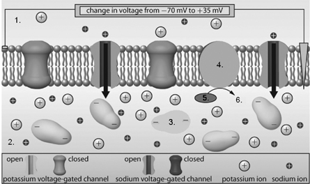
Adapted from Inquiry into Biology (Whitby, ON: McGraw-Hill Ryerson, 2007), BLM 11.1.9. Reproduced by permission.
SC 7. What is illustrated by number 1? Explain what is happening in number 1.
SC 8. What is shown by number 2? Explain what is happening to number 2 at this time.
SC 9. What is indicated by number 3? Identify one characteristic of number 3 and identify the function of number 3.
SC 10. What structure is shown by number 4? Explain its function.
SC 11. What are numbers 5 and 6? What is their function in nerve impulse transmission?
Self-Check Answers
SC 7. This shows a stimulated portion of a neuron membrane. The sodium gates are open and sodium ions are rushing to the inside of the neuron. The potassium gates are closed. The outside of the neuron is becoming negatively charged and the inside is becoming positively charged.
SC 8. The fluid inside the neuron (intracellular fluid) shows a high concentration of K+ ions and an increasing concentration of Na+ ions. The intracellular fluid is becoming positively charged.
SC 9. Large negatively charged protein particles, which are too big to move across the selectively permeable membrane, contribute to the voltage difference across the neuron membrane.
SC 10. This is a carrier protein embedded in the neuron membrane that functions in the sodium-potassium ion pump by using the energy of ATP to move three Na+ ions to the outside of the neuron and two K+ ions to the inside of the neuron. This maintains the difference in voltage across the membrane.
Repolarization of the Neuron
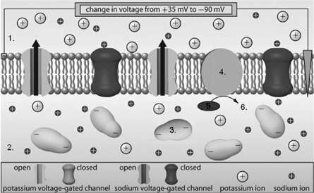
Adapted from Inquiry into Biology (Whitby, ON: McGraw-Hill Ryerson, 2007), BLM 11.1.9. Reproduced by permission.
SC 12. What is shown by number 1 in this diagram? How do you know this?
SC 13. What is happening in number 2 in this process?
SC 14. What is the function of structure 3?
SC 15. What functions does structure 4 serve in the process illustrated?
SC 16. What do 5 and 6 tell you about structure 4?
Self-Check Answers
SC 12. This shows a portion of the neural membrane undergoing repolarization. The sodium gates are closed and the potassium gates are open. K+ ions diffuse to the outside of the neuron and cause the outside to become positively charged.
SC 13. The fluid inside the neuron (intracellular fluid) is showing that Na+ ions are trapped on the inside of the neuron, and the inside of the neuron is becoming negatively charged.
SC 14. Large, negatively charged protein particles, which are too big to move across the selectively permeable membrane, contribute to the voltage difference across the neuron membrane.
SC 15. The sodium-potassium ion pump uses energy in the form of ATP to move three Na+ ions to the outside and two K+ ions to the inside to re-establish both the resting membrane potential and the correct distribution of ions.
If your answers are similar to the suggested answers, you are ready to submit an assignment to your teacher to show your understanding. If you do not understand the concepts, you need to access your teacher for further discussion before attempting the assignment.
 Module 1: Lesson 7 Assignment
Retrieve the copy of the Module 1: Lesson 7 Assignment that you saved to your computer earlier in this lesson. Complete Part 1 or Part 2, both of which deal with the concept of action potential. In Part 1 you may choose to analyze a graph and answer questions based on the graph. In Part 2 you may choose to summarize the events of the action potential. Save your completed assignment in your course folder. You will receive instructions about when to submit your assignment to your teacher later in this lesson.
1.49. Page 5
Module 1—The Nervous System
Myelin and Impulse Transmission
 Watch and Listen

© Jhaz Photography/shutterstock
In Lesson 3 you learned about the basic structures of the neuron. You discovered that some axons are wrapped in Schwann cells that produce a fatty material called myelin. Myelin acts as an insulator. In the following video clip, you will see the significance of insulation to communication in myelinated nerves versus unmyelinated nerves. Watch the following segment of “Nerve Impulse Conduction: Dentists Calms Your Nerves” before proceeding with the next section.
- "Bio Simulation: Myelinated vs. Nonmyelinated Neurons"
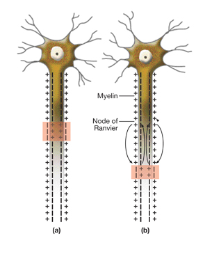
myelinated neuron: a neuron whose axon is wrapped by Schwann cells, which produces a myelin sheath
Myelinated neurons make up the white matter in the brain and the spinal cord and transmit nerve impulses very quickly.
unmyelinated neuron: a neuron that does not have Schwann cells and, therefore, lacks a myelin sheath
Unmyelinated neurons make up the grey matter in the brain and spinal cord and transmit nerve impulses much more slowly than myelinated neurons.
saltatory conduction: rapid transmission of a nerve impulse along an axon resulting from the action potential jumping from one node of Ranvier to another, skipping the myelinated regions of the membrane
In the video you saw that myelin is not present on all nerve cells. Myelinated neurons can conduct nerve impulses over 100 m/s, whereas unmyelinated neurons are much slower, with speeds of only 0.5 m/s. Even though the axon of the myelinated neuron is not in contact with the sodium-rich extracellular fluid outside the neuron, there is rapid communication. The nodes of Ranvier facilitate this rapid communication. In Lesson 3 you learned that the nodes of Ranvier lack myelin because they are between individual Schwann cells. Here, the axon is in contact with the extracellular fluid. The smooth wave-like nerve impulse that you studied in such detail in the previous lessons does not apply to myelinated neurons because action potentials can only occur at the nodes of Ranvier. Scientists found that the neural impulse “jumps” from one node to the next all along the axon in what is called saltatory conduction.
 Read
Read
For more detail on this process, read “Nerve Impulse” on pages 377 and 378 in the textbook. “Figure 11.16” on page 378 provides a good illustration of this process. Add this information to your course folder in a format that you prefer.

© 2008 Jupiterimages Corporation
Saltatory conduction is like a skillfully thrown rock skipping along a water surface, permitting a greatly increased speed of transmission. The speed of impulses is also increased in axons of large diameter. The fast-reacting giant axons of the squid that allow it to “jet-propel” itself away from danger are several millimeters in diameter, or 100 to 1000 times the diameter of a human axon. However, the squid’s giant axons conduct impulses at only about 30 m/s, which is far below your own capabilities.
Multiple Sclerosis
Watch and Listen
In Lesson 1 you were introduced to multiple sclerosis (MS) and some of the ways it affects communication in the nervous system.
Watch the segment called “Bio Discovery: Multiple Sclerosis” from “Electrochemical Control Systems in Humans: Regulating Physiological Processes.”
 Self-Check
SC 17. MS is a disorder capable of slowing down, or even stopping, impulse transmission. Prepare a paragraph outlining the relationship between myelinated nerve function and the symptoms of MS. Indicate the specific losses of myelinated nerve function caused by MS. After you check your answer, save it in your course folder for easy access when studying.
Self-Check Answers
SC 17. Multiple sclerosis (MS) is a disease of the white matter tissue of the central nervous system. The white matter is made up of myelinated nerve fibres, which are responsible for transmitting communication signals both within the CNS and between the CNS and the PNS. When the myelin sheaths of nerves of the CNS are damaged, nerve impulses are significantly slowed or even stopped. People with MS can experience partial or complete loss of any function that is controlled by or passes through the brain or the spinal cord. As such, this disease results in the weakening of the skeletal muscles. Note “Figure 11.17” on page 378 of your textbook, which shows lesions in the brain where the white matter has been destroyed. All myelinated motor neurons from the CNS to the skeletal muscles of the body are affected. This results in loss of muscle coordination and function.
1.50. Page 6
Module 1—The Nervous System
 Reflect and Connect
© Gina Sanders/shutterstock
In previous lessons you learned about the different parts of the nervous system and nerve transmission pathways. You also learned about sensory receptors that allowed you to hear the mosquito ring tone of a phone. Various lobes of the brain were involved in processing this information, and motor pathways led to responses. This lesson focused on the neuron, the type of cell that makes up the communication pathways of the nervous system. You discovered the special parts and functions of a neuron, and you considered how these structures facilitated communication and could even accelerate the rate of communication. This leads to an appreciation of the significance of myelination and an understanding of how symptoms of diseases such as MS indicate interruptions in communication.
 Module 1: Lesson 7 Assignment
Retrieve the copy of the Module 1: Lesson 7 Assignment that you saved to your computer earlier in this lesson. Complete Part 3—Reflect and Connect. In Part 3 you will have the opportunity to answer a series of questions based on data. This question is similar to the closed-response written question that you will write on the Diploma Exam at the end of this course. Save your completed assignment in your course folder. You will receive instructions about when to submit your assignment to your teacher later in this lesson.
Reflect on the Big Picture

© Dennis Sabo/shutterstock
In the Big Picture at the beginning of this module, you were introduced to unconscious and conscious communication in your nervous system. You now know that communication within or between any parts of your nervous system involve neurons. The Module Assessment involves research into Alzheimer’s disease—a disease involving major interruption in communication.
Research how Alzheimer’s affects the neuron and its ability to support communication. Take into particular consideration the consequences to communication when these affected neurons make up the brain. Store your research in your course folder. If you have any concerns about your research, contact your teacher.
Module 1: Lesson 7 Assignment
Submit your completed Module 1: Lesson 7 Assignment to your teacher for assessment.
1.51. Page 7
Module 1—The Nervous System
 Lesson Summary
In this challenging lesson you investigated the following focusing question:
- How does the structure of a neuron facilitate reception and transmission of a nerve impulse to the synaptic gap?
To answer this question, you explored the three major parts of nerve impulse transmission through a neuron:
- the resting or polarized state
- the action potential involving depolarization
- the re-establishment of the resting state, or repolarization
You investigated the importance of specific ions in communication. The distribution of the ions in the resting state produces a polarized membrane. In the resting state, the membrane of the neuron is impermeable to Na+ ions. The sodium-potassium ion exchange pump uses ATP to pump Na+ ions out of the neuron and K+ ions into the neuron. K+ ions diffuse through the selectively permeable membrane, but the negative ions attract them to keep most of the K+ ions inside the neuron. Thus, the positive charges dominate outside the neuron and the negative charges dominate the inside of the neuron. This creates the polarized state, with a voltage difference of about – 70 mV. Stimulation of a neuron causes depolarization, a shift in ion concentrations and electrical charges. You examined the mechanism of this shift.
When the neuron membrane becomes permeable to Na+ and the Na+ ions flood the inside of the neuron, the voltage difference becomes approximately + 40 mV and the permeability to Na+ is lost (sodium gates close). The K+ ions then leave the neuron and the resting potential of – 70 mV is restored. The sodium-potassium ion exchange pump works to bring the K+ back inside and kicks the Na+ out. This process takes only a millisecond or two and is called the refractory period. The neuron repolarizes in the refractory period and is then ready to be stimulated again. The nerve impulse is passed along the neuron in a wave of depolarization.
In this lesson you also learned that myelinated neurons conduct impulses much more quickly than nonmyelinated neurons because depolarization can only occur at the nodes of Ranvier. Action potentials at the node create enough current flow to activate the sodium gates in the next node, so the impulse “jumps” from node to node. Electrical current flow between nodes is instantaneous. Multiple sclerosis is an autoimmune disorder that causes the destruction of the myelin sheath, which ultimately slows down and stops the transmission of nerve impulses. This can result in blurred vision, loss of balance, and an inability of muscles to respond to commands from the brain, among other possible symptoms.
Lesson Glossary
Consult the glossary in the textbook for other definitions that you may need to complete your work.
action potential: the change in charge that occurs when the gates of the potassium ion channels close and the gates of the sodium ion channels open; a large depolarization event that is conducted along the membrane of a nerve cell or a muscle cell
all-or-none response: action that occurs either completely or not at all, such as the generation of an action potential by a neuron
depolarization: the loss or reduction of the negative resting membrane potential
hyperpolarization: the process of generating a membrane potential that is more negative than the normal resting membrane potential
membrane potential: a form of potential energy resulting from the separation of charges between the inside and the outside of a cell membrane; voltage across the cell membrane
myelinated neuron: a neuron whose axon is wrapped by Schwann cells, which produces a myelin sheath
Myelinated neurons make up the white matter of the brain and the spinal cord and transmit nerve impulses very quickly.
overshoot: the situation that results when more potassium ions leak out of the neuron than should because the potassium gates are slow to close; results in hyperpolarization
polarization: the process of generating a resting membrane potential averaging approximately – 70 mV
polarized membrane: the state of the cell membrane in an unstimulated neuron in which the inside of the neuron is negatively charged in comparison to the outside of the neuron; the resting state of a membrane averaging approximately – 70 mV
refractory period: the short time immediately after an action potential in which the neuron cannot respond to another stimulus; period of time it takes to re-establish the net positive charge on the outside of the neuron and the net negative charge on the inside of the neuron, where there are more sodium ions on the outside and more potassium ions on the inside of the neuron
repolarization: restoring the resting membrane potential (– 70 mV) from the depolarized state
resting membrane potential: the voltage that exists across a cell membrane during the resting state of an excitable cell, such as the neuron; averages around – 70 mV, but may range from – 50 to – 200 mV depending on the cell
saltatory conduction: rapid transmission of a nerve impulse along an axon resulting from the action potential jumping from one node of Ranvier to another, skipping the myelinated regions of the membrane
threshold potential: the smallest change in the membrane potential of a cell membrane that is needed to initiate an action potential; approximately – 55 mV
threshold stimulus: weakest possible stimulus that is needed to initiate a nerve impulse
unmyelinated neuron: a neuron that does not have Schwann cells and, therefore, lacks a myelin sheath
Unmyelinated neurons make up the grey matter of the brain and spinal cord and transmit nerve impulses much more slowly than myelinated neurons.
voltage: electrical potential difference across a membrane as measured by a voltmeter
1.52. Lesson 8
Module 1—The Nervous System
Lesson 8—Synaptic and Neuromuscular Transmission—Crossing the Divide
 Get Focused
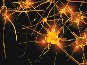
© Monika Wisniewska/shutterstock
You hear laughter from just behind you. You turn and your eyes meet those of the new girl you were introduced to earlier in the night. She smiles and asks, “Do you know the time?” What will you say? You are so nervous! Will a response even come out of your mouth? Thank goodness your nervous system is working, and you are able to stutter out the time.
Your nervous system is responsible for the communication between this person and yourself. Up to this point, you have learned about neurons and you are aware of the synaptic gaps, the tiny spaces between neurons. Communication usually requires some sort of “connection.” Think about what happens when the phone lines are down or your Internet connection doesn’t work. You know what it’s like when there are gaps in these communication pathways. When your phone and the Internet won’t work, there’s no “connection” to complete the communication pathway between you and the outside world!
In your nervous system, how do sights and sounds get communicated from neuron to neuron and across the synaptic gap between them? In most forms of communication, these incomplete connections would mean a communication “shut down,” but your nervous system is amazing! It has a unique way of completing the connection across the synaptic gap.
This lesson is about the events that occur in the synaptic gap. You will examine how neurons communicate with each other and with muscles. When scientists understood the events at the synaptic gap, technologies were developed to control and modify the communication of nerve transmissions. The events at the synaptic gap can be affected by many substances, such as coffee, energy drinks, alcohol, and anesthetics. They can also be affected by disorders such as Parkinson’s disease and Alzheimer’s disease.

left: © Dmitry Sunagatov/3409070/Fotolia; middle: © VR Photos/shutterstock; right: © VanHart/shutterstock
In this lesson you will investigate the following focusing questions:
- How does the anatomy and function of the synaptic gap and neuromuscular junction facilitate the transmission of nerve impulses between neurons and between neurons and effectors?
- How do chemicals taken into the body and disorders, such as Parkinson’s disease, compromise synaptic transmission?
 Module 1: Lesson 8 Assignment
Your teacher-marked Module 1: Lesson 8 Assignment requires that you complete and submit the following:
- Part 1—Drug Research
- Part 2—the Synaptic Gap
Download a copy of the Module 1: Lesson 8 Assignment to your computer now. You will receive further instructions about how to complete this assignment later in the lesson.
You must decide what to do with the questions that are not marked by the teacher.
Remember that these questions provide you with the practice and feedback that you need to successfully complete this course. You should respond to all the questions and place those answers in your course folder.
During this lesson, you will continue to develop your Module Assessment project by researching the relationship between Alzheimer’s disease and the events at the synaptic gap. As this is the last lesson in this module, you will have an opportunity to assemble all of your Module Assessment work and submit it for assessment. Your completed Module Assessment will be due at the end of this lesson.
You will also continue to add all your answers to questions, activities, and reflections to your course folder for later review and study. Research into Parkinson’s disease, curare, botulism, the characteristics of neurotransmitters, and the neural synapse will also be filed in your course folder.
Remember that further practice and application can be completed by completing questions in the textbook. The questions on pages 402 and 403 and pages 432 and 433 provide an excellent review of the material in this module. You may wish to answer these questions before you attempt the Module Assessment. You can access the answers to these questions and get feedback about your answers from your teacher. The Key and The Study Guide for Biology 30 provide you with many Diploma Exam-style multiple-choice, numerical-response, and written-response questions that will provide excellent review of the module and prove to be good preparation for the Diploma Exam.
1.53. Page 2
Module 1—The Nervous System
 Explore
Anatomy of the Synaptic Gap
 Read
Read
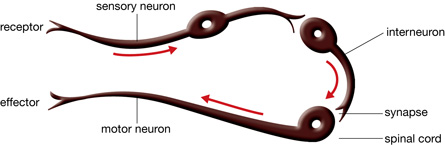
Your retinal receptor cells registered the new girl at school, the olfactory receptors detected the smell of her perfume, and the pressure receptors were activated by the contact of your handshake. The receptor cells converted this information to a nerve impulse, and information was on its way through a sensory neuron. But it came to a screeching halt at the end of the sensory neuron. Where does it go now? How does communication get to the next neuron? In the world of neurons, there’s a big gap to jump to the next neuron. To understand where the transmission goes next and the structures involved in the leap across the gap, study “Figure 11.18” on page 379 of your textbook.
synaptic knob: the tiny enlarged ending on an axon terminal
synaptic vesicle: a tiny membranous sac that, in this case, contains neurotransmitters
neurotransmitter: a chemical messenger released from the synaptic knob of a neuron at a synapse that diffuses across the synaptic cleft, binds to specially shaped protein receptors on the postsynaptic membrane, and stimulates the postsynaptic neuron
presynaptic membrane: the surface membrane surrounding the synaptic knob and facing the synaptic cleft
presynaptic neuron: the sending neuron
synaptic cleft: a tiny space separating the synaptic knob of a transmitting neuron from a receiving neuron or effector cell
postsynaptic neuron: the receiving neuron
postsynaptic membrane: the surface membrane on the dendrites of a receiving neuron across the synaptic cleft; contains receptor sites for neurotransmitters
Note the role of “shape” in controlling biological processes.
The synaptic knob is surrounded by the presynaptic membrane. The neuron that ends in the synaptic knob is called the presynaptic neuron. The synaptic cleft is the space between the presynaptic neuron and the next neuron, called the postsynaptic neuron. Remember that the receiving parts of the neuron are called the dendrites. Notice in the diagram on page 379 that the membrane surrounding the dendrite is called the postsynaptic membrane. Find the sodium ion channels in the postsynaptic membrane. Notice how these channels are only open when the neurotransmitter fits into a receptor. Note the role of shape in controlling biological processes.
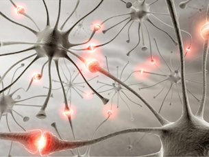
© iDesign/shutterstock
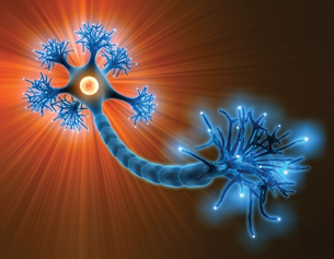
© iDesign/shutterstock

© David Hughes/shutterstock
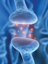
© Sebastian Kaulitzki/shutterstock
Crossing the Divide
Have you ever stood on a rock in a stream and wondered whether you could leap to the next rock? You think you can do it—you’re strong and co-ordinated—but if you fall, you could get very wet. To understand how a nerve transmission takes the “big leap” across the synaptic gap, study “Figure 11.18” on page 379 of the textbook and read "Signal Transmission Across a Synapse" on pages 378 to 380. Then follow up by looking at the animation showing synaptic transmission.
The diagram and readings illustrate that when the nerve impulse arrives at the synapse, it stimulates several reactions at the end of the axon. There is movement of the synaptic vesicles toward the presynaptic membrane, and then the vesicles fuse with the membrane. A neurotransmitter is released into the synaptic cleft, and it quickly diffuses across the synapse. Neurotransmitter molecules lock into receptor molecules on the postsynaptic membrane, causing the sodium gates to open and sodium ions from the synaptic cleft to rush into the postsynaptic neuron. You have also already learned that an inflow of sodium ions causes depolarization, and the start of an action potential. This now happens in the postsynaptic neuron (dendrite), resulting in a wave of depolarization through the postsynaptic neuron. The presynaptic and postsynaptic neurons have communicated a message across the gap via a type of chemical messenger called a neurotransmitter.
Summarize these events for your course folder as notes, a labelled diagram, a flow chart, or any format that you prefer.
 Watch and Listen
To visually explore synaptic transmission, you should choose to do one of the following:
- Watch the following segment of "Nerve Impulse Conduction: Dentists Calm Your Nerves." You may be required to enter a username and password to access the videos. Contact your teacher for this information.
- “Impulse Travel”
- “Impulse Travel”
- Watch the following segments of "Reflexes and Synaptic Transmission: Getting the Message Across."
- “Synaptic Transmission”
- “Bio Review: Synaptic Transmission”
As you watch any of the videos, answer the following questions. These questions will be an excellent reference for studying.
- Outline the reward pathway.
- What is a synapse?
- What happens at a synapse during synaptic transmission?
- What is a synaptic cleft? Is a synaptic cleft different from a synapse?
- What is a synaptic vesicle?
- What is the neurotransmitter in the reward pathway?
- Where are dopamine receptors located?
- What does the arriving nerve impulse do?
- What happens to the released neurotransmitter?
- Why is there a sucking sound in the video?
- What happens to the dopamine?
- How is the nerve impulse started in the second neuron?
- Identify two things that stop nerve impulse transmission.
- Does one neuron always synapse only with one other neuron? Suggest where this might be true.
1.54. Page 3
Module 1—The Nervous System
Reabsorption of Neurotransmitters
 Read
Read

© Robert Rozbora/shutterstock
Why don’t neurotransmitters continue to start new nerve impulses over and over again? This normally does not happen because the neurotransmitter is transported back into the presynaptic neuron, or it is broken down by enzymes and the fragments are moved back into the synaptic knob. For example, if the neurotransmitter is acetylcholine, the enzyme cholinesterase breaks it down and the fragments are reabsorbed into the synaptic knob. Scientists have created insecticides that bind up cholinesterase, causing acetylcholine to stay in the synapse and continually stimulate the muscles until they become fatigued and no longer contract. The insect dies because it can no longer contract muscles to get oxygen into its body. If the neurotransmitter is norepinephrine, it is reabsorbed directly into the presynaptic neuron. You can review what happens to the neurotransmitter by reading pages 380 and 382 in your textbook. Take notes as you read, and add this information to your course folder.
acetylcholine: one of the most common neurotransmitters of both the somatic nervous system and the parasympathetic nervous system
Acetylcholine functions by binding to receptors on the postsynaptic membrane and either depolarizing or hyperpolarizing the membrane.
cholinesterase: an enzyme necessary to decompose acetylcholine in the synaptic cleft so that the products (choline and ethanoic acid) can be reabsorbed by the presynaptic membrane; also called acetylcholinesterase
 Self-Check
To ensure that you understand the events that occur in synaptic transmission, answer the following questions. If you do not understand any part of synaptic transmission, or have any questions or concerns, discuss them with your teacher. Remember to phrase your answers in complete sentences using correct biological terminology. Consult the suggested answers for examples. Store your completed answers in your course folder.
SC 1. Summarize the events involved in impulse transmission, starting at the presynaptic neuron and ending at the postsynaptic neuron.
SC 2. Identify the function of neurotransmitters in the nervous system.
SC 3. Place the following events in the correct sequence.
- An action potential is initiated in the dendrite.
- Neurotransmitters are released into the synaptic cleft.
- Sodium gates are opened and sodium ions rush into the dendrite.
- A wave of depolarization arrives at the axon terminal.
SC 4. Deadly neurotoxins produced by some organisms, such as the rattlesnake and black widow spider, could have medical implications. One neurotoxin found in rattlesnake venom blocks receptors on postsynaptic neurons. Venom from the female black widow spider stimulates exocytosis of synaptic vesicles from neurons. Speculate how these neurotoxins might affect the body, and suggest a possible medical use for each of them.
© Miles Boyer/shutterstock
© Sly/401055/Fotolia
SC 5. Explain what happens to the neurotransmitters once an action potential has been started in the postsynaptic neuron.
Self-Check Answers
SC 1. The following events summarize, in correct sequence, the events involved in impulse transmission from the presynaptic neuron to the postsynaptic neuron.
- A wave of depolarization arrives at the axon terminal.
- Synaptic vesicles fuse with the presynaptic membrane.
- Neurotransmitters are released from the vesicles into the synaptic cleft.
- Neurotransmitters diffuse to the postsynaptic membranes and lock into complementary receptors.
- Sodium gates are opened and sodium ions rush into the postsynaptic neuron or dendrite of the second neuron.
- An action potential is initiated in the postsynaptic membrane of the dendrite.
- Neurotransmitters are decomposed by enzymes and reabsorbed by the presynaptic membrane.
SC 2. The function of neurotransmitters in synaptic transmission is to carry the neural signal from one neuron to another. Neurotransmitters can also carry the neural signal from a neuron to an effector, such as a muscle cell. Because the neurotransmitters can only be formed in the synaptic knob of the axon terminals, and neurotransmitter receptor sites are only located on the postsynaptic membrane, one-way impulse transmission is assured.
SC 3. The correct sequence of events is d, b, c, and a.
SC 4. One neurotoxin from rattlesnake venom blocks receptors on postsynaptic neurons. Venom from the female black widow spider stimulates exocytosis of synaptic vesicles from neurons. If a neurotoxin blocks receptors on postsynaptic neurons, as in the rattlesnake venom, it could prevent the binding of the neurotransmitter acetylcholine, thereby blocking the transmission of a nerve impulse to the muscle and causing muscle paralysis. If the impulse is a result of a negative stimulus, such as pain, blocking it could be effective in medical treatment. If a neurotoxin increases the release of neurotransmitters, as in the spider venom, it could also result in nerve and muscle fatigue and be useful in preventing transmission of pain impulses.
SC 5. If the neurotransmitter is acetylcholine, the enzyme cholinesterase (found in the synaptic cleft) decomposes the acetylcholine and the smaller fragments are reabsorbed by the presynaptic membrane. If the neurotransmitter is noradrenaline, it is reabsorbed directly.
SC 6. To review the anatomy of the synaptic gap and the functions of the structures, do this structures and functions drag-and-drop activity. Match the term with the number on the diagram, and match the function with the structure. Check your answers, and then file your work in your course folder.
1.55. Page 4
Module 1—The Nervous System
Neuromuscular Junctions
Now you know how messages are communicated from neuron to neuron through the nervous system. However, when you try to talk, how do the muscles in your mouth “get the message” from the neurons?

 Read
Read
When you read pages 380 to 382 in your textbook, you learned the details of the event that occur at the tiny gap located between the axon terminal and the muscle cell. You should choose to do one of the following to include this information in your course folder:
- Make summary notes for your course folder.
- Prepare a fully labelled diagram that outlines the process that occurs at the synaptic gap.
- Prepare a flow chart that outlines the events at the synaptic gap.
Make sure that your work addresses the following questions:
- What causes the synaptic vesicles to move toward the presynaptic membrane and fuse with it?
- What does the neurotransmitter that is released do?
- What does the fusion of the neurotransmitter, in this case acetylcholine, with the protein receptor do?
- What event does this initiate?
Please note that the figure incorrectly labels the sarcolemma as the neural membrane, when in fact the sarcolemma is a muscle cell membrane.
 Try This
neuromuscular junction: a tiny gap located between an axon terminal and a muscle cell
Interruption of neuromuscular transmission can have deadly effects on the body, particularly if the breathing muscles, such as the diaphragm, are affected. Research the following neurotoxins that act directly on neuromuscular junctions.
TR 1. The native tribes of South America apply a plant extract called curare to the tips of their arrows. Research how curare specifically affects neuromuscular junctions and why it is such an effective poison for the tribesmen.
TR 2. Botulism is a dreaded food poisoning that can be contracted by eating improperly canned foods. The neurotoxin involved in botulism is also the main ingredient in wrinkle-reduction injections called Botox. Research botulism to discover how the toxin specifically affects neuromuscular junctions. What is the result of this toxin on the body? Is there an antidote? How does botulism in the form of Botox injections reduce wrinkles?
1.56. Page 5
Module 1—The Nervous System
A Closer Look at Neurotransmitters
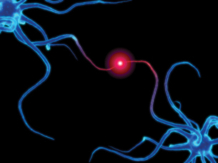
© Sebastian Kaulitzki/shutterstock
excitatory neurotransmitter: a neurotransmitter that promotes nerve impulse transmission in the postsynaptic membrane by opening sodium channels
inhibitory neurotransmitter: a neurotransmitter that hinders nerve impulse transmission in the postsynaptic neuron by hyperpolarizing it
Neurotransmitters are either excitatory or inhibitory at the synapse or at the neuromuscular junction. Sometimes the same neurotransmitter may be both inhibitory and excitatory, depending on the conditions in the body!
 Read
Read
dopamine: generally an excitatory neurotransmitter in the CNS
serotonin: an inhibitory neurotransmitter in the CNS
glutamate: an excitatory neurotransmitter in the CNS
GABA (gamma aminobutyric acid): an inhibitory neurotransmitter in the CNS
It is important to understand how neurotransmitters work. You should review pages 378 to 382 in your textbook. Begin reading at “Signal Transmission Across the Synapse.” Read up to the “Section Summary.”
By opening potassium channels or chloride channels in the postsynaptic membrane, inhibitory neurotransmitters make it harder to initiate depolarization in the second neuron because the neuron develops a much lower membrane potential—just like opening the windows on a winter day and letting the heat out makes it harder to warm up a room. Excitatory neurotransmitters open up sodium channels and make it easier to start depolarization because the membrane potential becomes less negative. These excitatory neurotransmitters act the same way as adding an electric heater to the room in order to offset the open window and make it easier to warm up the room. Acetylcholine and norepinephrine are just such excitatory neurotransmitters.
Acetylcholine is a common neurotransmitter in the somatic nervous system, and it is also found between the parasympathetic neurons of the autonomic nervous system. It is also found in some synapses of the brain. Norepinephrine (also called noradrenalin) is an important neurotransmitter in the sympathetic nervous system, and is also found in some synapses in the brain. Review pages 378 to 382 in your textbook and study the table below to learn more about some important neurotransmitters and their characteristics.
CHARACTERISTICS OF SELECTED NEUROTRANSMITTERS
Neurotransmitter |
Secretion Location |
Type |
Effects of Abnormal Production |
acetylcholine |
CNS, PNS, neuromuscular junctions |
mostly excitatory |
|
norepinephrine |
CNS, PNS, sympathetic neurons |
excitatory in sympathetic neurons |
|
dopamine |
CNS |
mostly excitatory |
|
serotonin |
CNS |
generally inhibitory |
|
glutamate |
CNS |
excitatory |
|
GABA (gamma aminobutyric acid) |
CNS |
inhibitory |
|
endorphins |
CNS |
inhibitory |
|
 Try This
Choose one of the following Try This activities.
TR 3. Using your textbook, the Internet, and any other sources that may be available to you, research what happens in the body when acetylcholine, norepinephrine, dopamine, serotonin, glutamate, GABA, or endorphins are not produced in appropriate amounts in the body. Add this new information to a table like the one above.
OR
TR 4. You may choose to do questions 6, 7, and 8 on page 384 of the textbook. Check your answers with your teacher.
 Discuss
The inability of the body to produce appropriate amounts of a neurotransmitter can result in serious consequences for the body. Michael J. Fox and Mohammed Ali are two famous people who have been afflicted with Parkinson’s disease. This disease seriously interrupts communication in the nervous system. Research the following questions about Parkinson’s disease. Post your research on the discussion board, or build a wiki with a group of students. If you are building a wiki, you can split up the questions for different group members to complete. After reviewing the shared research and based on the feedback from your peers, make any necessary modifications to your work. You may choose to further discuss your research with your teacher. Save your research in your course folder.
- Define Parkinson’s disease.
- List the symptoms that are associated with Parkinson’s disease, and explain these symptoms based on your understanding of this lesson.
- Outline the causes of Parkinson’s disease.
- Describe two possible treatments for someone with Parkinson’s disease.
- Briefly explain what a stem cell is, and how it can be used to treat Parkinson’s disease.
- Outline the societal and technological issues associated with using embryonic stem cell transplants to treat Parkinson’s disease.
 Self-Check
Check your understanding of the concept of neurotransmitters by answering the following questions in full sentences. If at any point you require more help, consult with your teacher.
SC 7. Compare the effects of excitatory and inhibitory neurotransmitters on the postsynaptic membrane.
SC 8. Explain the relationship between acetylcholine and cholinesterase.
SC 9. Parkinson’s disease is associated with which neurotransmitter? Identify which area of the brain is affected by this disease and the evident symptom that results.
Self-Check Answers
SC 7. Both excitatory and inhibitory neurotransmitters affect the transmission of the nerve impulse across the synapse. (Similarity) Excitatory neurotransmitters generally make it easier to initiate depolarization in the dendrites of the postsynaptic neuron by opening sodium channels in the postsynaptic membrane and allowing sodium ions to diffuse into the postsynaptic neuron, thus initiating the action potential in the second neuron. Inhibitory neurotransmitters make it more difficult to initiate depolarization in the postsynaptic neuron by either opening potassium channels and allowing more potassium ions to leak out of the neuron (hyperpolarizing it), or by opening chloride channels and allowing negative chloride ions to leak into the neuron, therefore making the neuron more negative than its normal resting potential of – 60 or – 70 mV.
SC 8. After acetylcholine has opened sodium channels in the postsynaptic membrane and initiated depolarization in the postsynaptic neuron, it is decomposed by the enzyme cholinesterase, and the products are transported into the presynaptic neuron so that they can be resynthesized into acetylcholine. If cholinesterase is unable to catalyze the decomposition of acetylcholine, the acetylcholine remains in the synaptic cleft and in the receptors and stimulates the postsynaptic neuron continuously, which may cause muscle fatigue.
SC 9. Parkinson’s disease is associated with the neurotransmitter dopamine. Parkinson’s disease occurs when the nerve cells in the part of the brain that controls muscle movement are gradually destroyed and the neurons can no longer produce dopamine so that muscles can be co-ordinated. Consequently, some of the initial symptoms include muscle stiffness and difficulty bending the arms or legs; an unstable, stooped, or slumped-over posture; loss of balance; a slow, shuffling walk; varying degrees of shaking or muscle tremors; reduced ability to show facial expressions; slow speech; and loss of fine motor skills, such as the inability to write.
1.57. Page 6
Module 1—The Nervous System
The Effect of Drugs on Neurons and Synapses
A drug is a substance that changes the way the body functions. Most drugs, whether they are legal or illegal, affect the neural synapses by either enhancing or decreasing the action of a neurotransmitter. They do this by affecting the vesicles, the receptor proteins, the ion gates in the postsynaptic membrane, or the reabsorption of neurotransmitters.
 Read
Read
To understand how drugs interrupt communication, read page 383 of your textbook.
 Try This
Choose one of the following Try This activities.
TR 5. Investigate in greater detail how drugs affect neurons and synapses in “Drugs Alter the Brain’s Reward Pathway.” Type the following address into a new browser window: learn.genetics.utah.edu/units/addiction/drugs/. Explore one of the following articles at the website:
- “Explore! Drugs of Abuse”
- “The Physiology of the High”
- “Hardwiring an Addict”
- “Changes Last Long After Use”
- “How PET Scans Can Measure Brain Activity”
- “Death By Overdose”
OR
TR 6. Type the following address into a new browser window: learn.genetics.utah.edu/units/addiction/drugs/. Click on “Mouse Party” in “Drugs Alter the Brain’s Reward Pathway.” As you study the effects of the various drugs on mice, reflect on the questions outlined for each mouse. Based on your knowledge, formulate a hypothesis to explain the results you observe.
You will be able to use this information when you prepare your lesson assessment.
 Module 1: Lesson 8 Assignment
Retrieve the copy of the Module 1: Lesson 8 Assignment that you saved to your computer earlier in this lesson. Complete Part 1—Drug Research, where you are asked to research three different drugs. Save your completed assignment in your course folder. You will receive instructions about when to submit your assignment to your teacher later in this lesson.
1.58. Page 7
Module 1—The Nervous System
 Self-Check
Synaptic and neuromuscular transmitters are important and often challenging concepts to master. To ensure your understanding, complete the following Self-Check questions. The Self-Check is optional, but it is an excellent review and preparation for your lesson assignment that you will submit for assessment.
Use the following information to answer the next two questions.
| Avoiding a Collision
You are driving down a highway at night when a deer jumps in front of your vehicle. You slam on the brakes and avoid a collision. |
SC 10. Which row below best describes the initial reaction of your autonomic nervous system to this situation?
Row |
System Involved |
Neurotransmitter |
Response |
A. |
sympathetic nervous system |
norepinephrine | heart rate increases; pupils of eyes dilate |
B. |
parasympathetic nervous system |
acetylcholine | heart rate increases; pupils of eyes dilate |
C. |
sympathetic nervous system |
acetylcholine | heart rate increases; pupils of eyes constrict |
D. |
parasympathetic nervous system |
aorepinephrine | heart rate decreases; pupils of eyes constrict |
SC 11. Which row below best describes the reaction of your autonomic nervous system several minutes after the incident?
Row |
System Involved |
Neurotransmitter |
Response |
A. |
sympathetic nervous system |
norepinephrine | heart rate increases; pupils of eyes dilate |
B. |
parasympathetic nervous system |
norepinephrine | heart rate increases; pupils of eyes dilate |
C. |
sympathetic nervous system |
acetylcholine | heart rate decreases; pupils of eyes constrict |
D. |
parasympathetic nervous system |
acetylcholine | heart rate decreases; pupils of eyes constrict |
Use the following information to answer the next question.
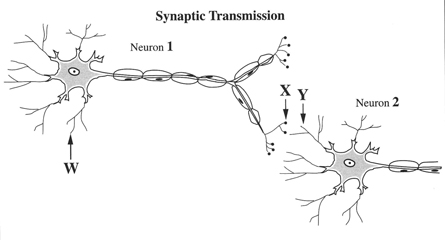
SC 12. Destruction of the synaptic vesicles of Neuron 1 will
- block the nerve impulse at W
- cause X to be constantly stimulated
- prevent depolarization from occurring at Y
- result in the action of cholinesterase in Neuron 2
Use the following information to answer the next question.
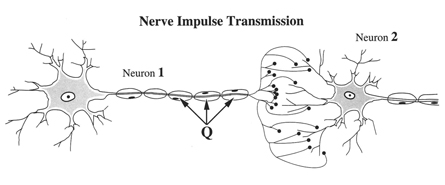
SC 13. If the structures labelled Q were absent, what effect on neural transmission would be expected?
- The axon would not release acetylcholine.
- The axon would not become depolarized.
- The speed of transmission would be reduced.
- No action potential would arrive to release neurotransmitter.
Use the following information to answer the next question.
| The disease myasthenia gravis causes a person to experience muscular weakness because of the failure of neuromuscular junctions to transmit signals from nerve fibres to muscle fibres. The weakness is due to a reduced sensitivity to acetylcholine, which is necessary to stimulate the muscle fibre. People suffering from this disease are often treated with neostigmine, an anticholinesterase drug, which can result in some normal muscular activity within minutes.—Guyton and Hall, 1996 |
SC 14. Neostigmine is effective in treating this disease because it
- binds with cholinesterase to form acetylcholine
- binds with cholinesterase to increase acetylcholine production
- reduces the amount of active cholinesterase, thereby increasing the amount of acetylcholine available to stimulate muscle contraction
- increases the amount of active cholinesterase, thereby increasing the amount of acetylcholine available to stimulate muscle contraction
Use the following information to answer the next question.
Observations About a Synapse and Synaptic Transmission
|
SC 15. The assumption that axon terminals contain a limited amount of neurotransmitter could account for observation
- 1
- 2
- 3
- 4
Use the following information to answer the next question.
Alternative medicine, such as aromatherapy, is becoming increasingly popular in western society. Aromatherapy uses natural oils and plant extracts. The scents of the oils and extracts are inhaled or the fragrant oils are massaged into the skin. Proponents of aromatherapy hypothesize that odours affect the brain and its release of neurochemicals. These neurochemicals may then relieve pain.
Hypothesized Steps in Aromatherapy Action
|
Numerical Response
SC 16. If it is assumed that the hypothesis is correct, the order in which the steps above would occur to result in pain relief in a person having just inhaled the scent from an aromatherapy oil or extract is ______, ______, ______, and ______.
SC 17. While working in a lab, you accidentally mixed some unknown chemicals together and a vapour was produced, which diffused throughout the lab. Soon the lab animals in the room collapsed, unable to move. You made a quick examination of the animals and found their musculature to be very loose and relaxed. You concluded that the vapour became internalized in the animals by way of the lungs to the blood, which then distributed the vapour throughout the animals’ bodies. The symptoms shown by the lab animals may lead you to conclude that the vapour could have
- stimulated the action of the enzyme cholinesterase
- inhibited the production of acetylcholine by the axon endings of neurons
- inhibited the formation of synaptic transmission chemical produced by the dendrites
- stimulated the axon endings to secrete large amounts of acetylcholine
Self-Check Answers
SC 10. A
SC 11. D
SC 12. C
SC 13. C
SC 14. C
SC 15. C
SC 16. 2, 1, 4, 3
SC 17. B
1.59. Page 8
Module 1—The Nervous System
 Reflect and Connect
In this lesson you reviewed the function and physiology of neurotransmitters. In this part of your assignment, you will consider a diagram of a sensory neuron and a motor neuron and answer a series of questions about the synapse and the relationship between these two neurons. These questions are similar to the closed written-response question on the Diploma Exam.
 Module 1: Lesson 8 Assignment
Retrieve the copy of the Module 1: Lesson 8 Assignment that you saved to your computer earlier in this lesson. Complete Part 2—The Synaptic Gap. Save your completed assignment in your course folder. You will receive instructions about when to submit your assignment to your teacher later in this lesson.
 Reflect on the Big Picture
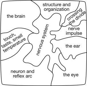
From the Big Picture scenario, your rapidly beating heart, your increased breathing rate, and the simple act of walking across the room take on a whole new meaning. In this lesson you examined how the information communicated through neurons can then be transmitted across the gap between neurons or between a neuron and a muscle. You examined the various types of neurotransmitters found naturally in the body, and you should now understand how they can speed up or slow down communication. You also looked at the effects of drugs on the synaptic gap.
Your Module Assessment project about Alzheimer’s disease will be completed with this lesson. Based on your understandings from this lesson, you should research any relationship between the symptoms of the disease and abnormal events that occur at the synaptic gap. Does the synaptic gap play any role in the interruption of communication associated with Alzheimer’s disease? Store your research findings in your course folder in preparation of your final work on this project.
Module 1: Lesson 8 Assignment
Submit your completed Module 1: Lesson 8 Assignment and your Module Assessment to your teacher for assessment.
1.60. Page 9
Module 1—The Nervous System
 Lesson Summary
In this lesson, you have explored the following focusing questions:
- How does the anatomy and function of the synaptic gap and neuromuscular junction facilitate the transmission of nerve impulses between neurons and between neurons and effectors?
- How do chemicals taken into the body and disorders, such as Parkinson’s disease, compromise synaptic transmission?
The parts of the nervous system—neurons, neural pathways, receptors, effectors, and synapses—all fit together like the pieces of a puzzle. If one piece fails to fit, the system doesn’t work right. Disorders, such as Parkinson’s disease, and chemical substances, such as drugs, can interrupt the function of the system.
Lesson Glossary
Consult the glossary in the textbook for other definitions that you may need to complete your work.
acetylcholine: one of the most common neurotransmitters of both the somatic nervous system and the parasympathetic nervous system
Acetylcholine functions by binding to receptors on the postsynaptic membrane and either depolarizing or hyperpolarizing the membrane.
cholinesterase: an enzyme necessary to decompose acetylcholine in the synaptic cleft so that the products (choline and ethanoic acid) can be reabsorbed by the presynaptic membrane; also called acetylcholinesterase
dopamine: generally an excitatory neurotransmitter in the CNS
excitatory neurotransmitter: a neurotransmitter that promotes nerve impulse transmission in the postsynaptic membrane by opening sodium channels
GABA (gamma aminobutyric acid): an inhibitory neurotransmitter in the CNS
glutamate: an excitatory neurotransmitter in the CNS
inhibitory neurotransmitter: a neurotransmitter that hinders nerve impulse transmission in the postsynaptic neuron by hyperpolarizing it
neuromuscular junction: a tiny gap located between an axon terminal and a muscle cell
neurotransmitter: a chemical messenger released from the synaptic knob of a neuron at a synapse that diffuses across the synaptic cleft, binds to specially shaped protein receptors on the postsynaptic membrane, and stimulates the postsynaptic neuron
norepinephrine: a neurotransmitter released by sympathetic neurons of the autonomic system to produce an excitatory effect on target muscles; also called noradrenalin
postsynaptic membrane:the surface membrane on the dendrites of a receiving neuron across the synaptic cleft; contains receptor sites for neurotransmitters
Note the role of “shape” in controlling biological processes.
postsynaptic neuron: the receiving neuron
presynaptic membrane: the surface membrane surrounding the synaptic knob and facing the synaptic cleft
presynaptic neuron: the sending neuron
serotonin: an inhibitory neurotransmitter in the CNS
synapse (also synaptic cleft and synaptic gap): the location or junction in a neural pathway where one neuron communicates with another neuron; a tiny gap between the synaptic terminal of an axon and the signal receiving dendrite or cell body of another neuron or an effector, such as a muscle
synaptic cleft: a tiny space separating the synaptic knob of a transmitting neuron from a receiving neuron or effector cell
synaptic knob: the tiny enlarged ending on an axon terminal
synaptic vesicle: a tiny membranous sac that, in this case, contains neurotransmitters
1.61. Module Summary/Assessment
Module 1—The Nervous System
 Module Summary
In this module you were asked the following inquiry question:
- How does the nervous system communicate with the body and maintain homeostasis?
You investigated how various sensory cells and organs help you to see a person, hear their laughter from across the room, and detect his or her scent. You explored how these sensations were transmitted electrochemically between neurons to specific areas of the brain so that they could be interpreted. You discovered how your brain was able to send nerve impulses to your leg muscles so that you could walk in the direction of the person on the other side of the room. These communications were conscious. Other parts of the brain, such as the medulla oblongata, sent out unconscious messages via the sympathetic nervous system. These messages altered your heart rate and caused your hands to become clammy. After that initial “hello,” your parasympathetic nervous system slowed the heart down and returned your body to homeostasis. All of these different parts of the nervous system communicate with the body and work towards sustaining homeostasis. Some of the parts are related to other systems, while others parts function independently.
Module 1 provided an opportunity to investigate how several disorders, such as multiple sclerosis, Alzheimer’s disease, and Parkinson’s disease, disrupt communication in the nervous system, cause a loss of homeostasis, and require corrective technologies to attempt to bring the system back to homeostasis.
To review and summarize the concepts of Module 1, you may wish to complete the Module 1 Concept Organizer. You may have already saved a copy of this document when you first encountered it in the Big Picture section, or you may download it now. It is an outline of the lessons that contains the focusing questions for Module 1. Use the focusing questions to fill in this concept organizer with the ideas that you mastered in each lesson. You can use keywords, point form, or any amount of detail that meets your needs. You may choose to work from the file on your computer, print the document and work from the paper copy, or copy the outline onto a large sheet of poster paper. This is a great tool to review and use for study purposes.
For this module a completed version of the concept organizer is provided for review purposes. As you work through the course, you will be provided with an outline Concept Organizer. Use this outline to complete your own version to meet your study needs.
 Module Assessment
Before you begin the Module Assessment, you may choose to complete the review questions on pages 402 and 403 and pages 432 and 433 of your textbook. Your teacher will help you with any questions that you might have and give you feedback on your responses.
For this Module Assessment, you may choose to create a pamphlet, slideshow presentation, website, TV or podcast infomercial recording or transcripts, annotated collage, or other approved form of media to provide information about Alzheimer’s disease. With each lesson you completed in Module 1, you acquired new knowledge and information about the nervous system as it relates to Alzheimer’s disease. You will need to refer to the information you saved in your course folder as preparation work for this project.
You should explore the following questions in your Module Assessment project:
- What is Alzheimer’s disease?
- Who is susceptible to the disease?
- What are the symptoms?
- What parts of the nervous system are affected?
- What happens to the neurons?
- Is impulse transmission affected?
- Is synaptic transmission compromised?
- What are some technologies that are used to investigate Alzheimer’s disease?
- How is Alzheimer’s disease treated?
- Are there any new lines of research?
You will post your work on the discussion board and discuss it with other students. What did they like about your project? What areas need improvement? What did you leave out?
Your work will be submitted to your teacher for marking.
Rubric
| 5 | means you have completed every part of the question with detailed, well-thought-out answers |
| 4 | means that you have completed every part of the question, but your answers are brief and lack the detail necessary for a well-thought-out answer |
| 3 | means that you have omitted some parts of the question |
| 2 | means very little information has been presented and what has is either incorrect or missing important detail |
As you prepare your response, you may wish to indicate your progress by checking the box in front of each bullet. This will help you evaluate and reflect on your work before submitting your response to your teacher.
Score |
Scoring Criteria – The student … |
5 Excellent |
|
4 |
|
3 |
|
2 |
|
1 |
|
Note: A mark of “Insufficient” may be assigned to projects that do not contain a discernable attempt to address the assignment or are too brief to assess.
1.62. Module Glossary
Module 1—The Nervous System
Module Glossary
Consult the glossary in the textbook for other definitions that you may need to complete your work.
accommodation: the process of changing the shape of the lens from round and fat to thin and flat, and vice versa, so that light can be focused on the retina to accommodate vision of objects near and far away
acetylcholine: one of the most common neurotransmitters of both the somatic nervous system and the parasympathetic nervous system
Acetylcholine functions by binding to receptors on the postsynaptic membrane and either depolarizing or hyperpolarizing the membrane.
action potential: the change in charge that occurs when the gates of the potassium ion channels close and the gates of the sodium ion channels open; a large depolarization event that is conducted along the membrane of a nerve cell or a muscle cell
adaptation: the process by which the iris adjusts the diameter of the pupil based on light conditions, thus controlling the amount of light that enters the eye and strikes the retina
all-or-none response: action that occurs either completely or not at all, such as the generation of an action potential by a neuron
amplify: to increase the amplitude or the extent of a vibration; see intensity
anterior chamber: the space in front of the iris and behind the cornea that is filled with aqueous humour
aqueous humour: a clear, watery fluid in the anterior chamber of the eye that maintains the shape of the cornea and provides oxygen and nutrients for the surrounding cells, including those of the lens and the cornea
astigmatism: an uneven curvature of the cornea or lens, resulting in uneven focusing, which results in poor vision
auditory canal: a short channel that funnels sound waves from outside the ear to the tympanum and amplifies sounds
auditory nerve: a nerve composed of sensory fibres from the organ of Corti, the vestibular apparatus that conducts impulses to the temporal lobe of the cerebrum
autonomic nervous system (ANS): a division of the peripheral nervous system that conducts nerve impulses to cardiac and smooth muscles, as well as to glands; may also be called the involuntary motor system
axon: the long extension that emerges from the cell body and conducts the nerve impulse away from the cell body
The axon may be up to 1 m long in motor neurons.
axon terminal: numerous endings found at the end of an axon
Axon terminals are enlarged into knobs that are specialized for producing, storing, and releasing neurotransmitters.
basilar membrane: one of two parallel membranes that comprise the organ of Corti in the inner ear
Hair cells are attached to the basilar membrane.
bipolar cells: specialized sensory nerve cells located in the retina that are stimulated by either rods or cones
Cones mostly have a one-to-one ratio with bipolar cells, whereas several rod cells may communicate with one bipolar cell.
blind spot: the area at the back of the eyeball that is deficient in rods and cones; the area where the sensory fibres come together to form the optic nerve
cataract: a cloudy or grey-white area on the lens caused by deterioration of the protein composing the lens
Cataracts prevent the passing of light to the photoreceptors of the retina.
cell body: the part of a neuron that contains the nucleus and other cell organelles for carrying out the metabolic reactions of the nerve cell; relays the nerve impulse from the dendrites to the axon
central nervous system (CNS): the part of the nervous system that includes the brain and spinal cord
chemoreceptor: a sensory receptor that transmits information about the solute concentration in a solution or about individual kinds of molecules in solution
cholinesterase: an enzyme necessary to decompose acetylcholine in the synaptic cleft so that the products (choline and ethanoic acid) can be reabsorbed by the presynaptic membrane; also called acetylcholinesterase
choroid: the middle layer of the eyeball that lies between the sclera and retina and is highly vascular and heavily pigmented
The choroid absorbs stray light rays not detected by the photoreceptors of the retina.
ciliary muscle: a ring of muscle behind the iris that is attached to the lens by suspensory ligaments and is involved in changing the shape of the lens
cochlea: contains the organ of Corti, which functions to convert the mechanical energy of sound waves into a nerve impulse
colourblindness: an x-linked inherited disorder that results in nonfunctional or deficient cone function; inability to see certain colours such as red, green, or blue
cones: one of two types of photoreceptors in the retina of the eye that is sensitive to different wavelengths of light and are, thus, responsible for distinguishing colour
There are three types of cones: one sensitive to red light, one sensitive to blue light, and one sensitive to green light. The cones are responsible for acute vision, or distinguishing detail.
cornea: a transparent portion of the sclera (located at the front of the eye) that allows light to enter the eye and, in the process, refracts or bends the light rays so that they can be focused on the retina
dendrite: a short, branching terminal of a neuron that receives input from other neurons or sensory receptors and transmits a nerve impulse toward the cell body
depolarization: the loss or reduction of the negative resting membrane potential
depth perception: the ability to see in three dimensions
dopamine: generally an excitatory neurotransmitter in the CNS
dynamic balance (rotational equilibrium): balance resulting when the head and body are moved or rotated
effector: one of the three types of muscle or a gland that responds to a nerve impulse
Eustachian tube: a tiny passageway extending from the middle ear to the throat (pharynx); a passageway through which viruses and bacteria can travel from the nose and mouth, resulting in an inner ear infection
The Eustachian tube plays an important role in equalizing air pressure on both sides of the tympanum.
excitatory neurotransmitter: a neurotransmitter that promotes nerve impulse transmission in the postsynaptic membrane by opening sodium channels
fovea centralis: an area of the retina that is located directly behind the centre of the lens and has a very high concentration of cones, which makes this part of the eye responsible for great visual acuity
frequency: number of wavelengths per given time; number of waves that pass a given point in a given time
GABA (gamma aminobutyric acid): an inhibitory neurotransmitter in the CNS
ganglion cells: special sensory neurons that communicate with bipolar cells in the retina to transmit a nerve impulse to the brain
These cells have long axons that converge at the back of the eye to form the optic nerve.
glaucoma: a disorder caused by the malfunction of ducts that drain excess aqueous humour from the anterior chamber
The resulting pressure created by excess aqueous humour ruptures delicate blood vessels in the eye and causes deterioration of cells in the eye due to lack of nutrients. This can result in blindness if left untreated.
glutamate: an excitatory neurotransmitter in the CNS
hair cells: sensory mechanoreceptors attached to the basilar membrane in the organ of Corti
homeostasis: a state of body equilibrium or a stable internal environment of the body
hyperopia: farsightedness, or the inability to focus objects that are close, caused by an eyeball that is too short, which causes light to be focused behind the retina
hyperpolarization: the process of generating a membrane potential that is more negative than the normal resting membrane potential
incus (anvil): the middle in the sequence of the three ossicles located in the middle ear; connected to the malleus and the third ossicle (stapes); part of a lever system that amplifies sound waves
inhibitory neurotransmitter: a neurotransmitter that hinders nerve impulse transmission in the postsynaptic neuron by hyperpolarizing it
inner ear: one of the three regions of the ear that is located deepest into the head and consists of a fluid-filled chamber containing the semicircular canals, the utricle and saccule, and the cochlea
intensity: measurement of the difference between compressed and rarefied areas of a sound wave used in physics; corresponds to amplitude of a wave
interneuron: a type of nerve cell found either in the brain or spinal cord that transmits nerve impulses from sensory neurons within the various parts of the brain or to motor neurons
iodopsin: the general name of any of the three visual pigments found in cone cells that is stimulated by light to initiate a nerve impulse
loudness: subjective interpretation of sound intensity
malleus (hammer): the first in the sequence of the three ossicles located in the middle ear; connected to the tympanum and the second ossicle (incus); part of a lever system that amplifies sound waves
mechanoreceptor: a sensory receptor that detects physical deformations in the body’s environment associated with pressure, touch, stretch, motion, and sound
membrane potential: a form of potential energy resulting from the separation of charges between the inside and the outside of a cell membrane; voltage across the cell membrane
middle ear: one of the three main regions of the ear, which begins just past the tympanum and consists of a chamber containing three tiny bones (ossicles) called the malleus (hammer), the incus (anvil), and the stapes (stirrup); leads into a minute opening called the Eustachian tube
motor neuron: a type of nerve cell that transmits nerve impulses toward an effector, which can be a muscle or a gland
myelinated neuron: a neuron whose axon is wrapped by Schwann cells, which produces a myelin sheath
Myelinated neurons make up the white matter of the brain and the spinal cord and transmit nerve impulses very quickly.
myelin sheath: a fatty insulating layer that surrounds axons and greatly increases the rate of impulse transmission and maintains the strength of the impulse by preventing the loss of ions along the length of the axon
myopia: nearsightedness, or the inability to focus objects that are far away, caused by an eyeball that is elongated, which causes light to be focused in front of retina rather than directly on it
nerve: a message pathway of the nervous system; made up of many neurons grouped into bundles and surrounded by protective tissue
There are 12 pairs of cranial nerves that insert into the brain and 31 pairs of spinal nerves that emanate from the spinal cord.
nervous system: an elaborate communication system that receives input; processes, integrates, and stores information; and triggers muscle contraction or glandular secretion
neuromuscular junction: a tiny gap located between an axon terminal and a muscle cell
neuron: a cell in the nervous system that generates and transmits nerve impulses; consists of dendrites, a cell body containing the nucleus, and an axon that may or may not have a myelin sheath
neurotransmitter: a chemical messenger released from the synaptic knob of a neuron at a synapse that diffuses across the synaptic cleft, binds to specially shaped protein receptors on the postsynaptic membrane, and stimulates the postsynaptic neuron
node of Ranvier: a tiny gap in the myelin sheath surrounding the axon of myelinated neurons
Nerve impulse transmission occurs between nodes of Ranvier in what is called salutatory conduction, which causes the increase in the speed of impulse transmission.
norepinephrine: a neurotransmitter released by sympathetic neurons of the autonomic system to produce an excitatory effect on target muscles; also called noradrenalin
occipital lobe: one of the four lobes of the cerebrum that receives and analyzes visual information that is sent to association centres for recognition of what is being seen
olfactory (receptor) cell: a neuron located in the olfactory epithelium that is specialized to receive chemical stimuli and to initiate a nerve impulse
olfactory epithelium: a patch of tissue located in the upper part of the nasal cavity that contains mucous-secreting cells and olfactory cells, or smell cells, that can detect different smells; a sense organ for smell
opsin: a protein that is the result of the decomposition of rhodopsin
optic nerve: a collection of sensory neurons that carries sensory information from the photoreceptors to the brain
organ of Corti: a structure of the inner ear that contains hair cells that detect vibrations in the fluid of the inner ear and initiates a nerve impulse that is transmitted to the auditory nerve
osmoreceptor: a sensory receptor that detects changes in osmotic pressure, pressure due to water movement
ossicles: three tiny bones located in the middle ear that are connected to each other, to the tympanum, and to the oval window; involved in amplifying sound waves
otolith: a tiny particle of calcium carbonate found in the utricle and saccule that contacts the hair cells of these structures and stimulates them
outer ear: one of the three main regions of the ear, which consists of the pinna and the auditory canal and ends at the tympanum or eardrum
oval window: a membrane-covered opening located between the chamber of the middle ear and the wall of the inner ear
The stapes (stirrup) is attached to it and transmits sound waves by it to the inner ear.
overshoot: the situation that results when more potassium ions leak out of the neuron than should because the potassium gates are slow to close; results in hyperpolarization
parasympathetic nervous system: the division of the autonomic nervous system that oversees digestion, elimination, and glandular function; often works opposite the sympathetic nervous system to bring the body back to normal
perception: the interpretation of sensory information by the cerebral cortex
peripheral nervous system (PNS): the portion of the nervous system consisting of nerves and ganglia (collections of nerve cell bodies) that are outside the brain and spinal cord
photoreceptor: a sensory receptor that responds to light stimuli, allowing people to see images and colours
pinna: the outer flap of the ear that is composed of skin and cartilage and shaped so that it enhances sound vibrations and funnels them into the auditory canal
pitch: corresponds to the frequency of a sound wave
polarization: the process of generating a resting membrane potential averaging approximately – 70 mV
polarized membrane: the state of the cell membrane in an unstimulated neuron in which the inside of the neuron is negatively charged in comparison to the outside of the neuron; the resting state of a membrane averaging approximately – 70 mV
postsynaptic membrane: the surface membrane on the dendrites of a receiving neuron across the synaptic cleft; contains receptor sites for neurotransmitters
Note the role of “shape” in controlling biological processes.
postsynaptic neuron: the receiving neuron
presynaptic membrane: the surface membrane surrounding the synaptic knob and facing the synaptic cleft
presynaptic neuron: the sending neuron
reflex: an inborn, unlearned behaviour that results from the stimulation of a special neural pathway called the reflex arc
reflex arc: an involuntary neural pathway that consists of a sensory receptor, a sensory neuron, a control centre that can be either the brain or spinal cord, a motor neuron, and an effector that results in a reflex behaviour that usually has survival value
refract: to bend light as it passes through a substance with a different desnsity
refractory period: the short time immediately after an action potential in which the neuron cannot respond to another stimulus; period of time it takes to re-establish the net positive charge on the outside of the neuron and the net negative charge on the inside of the neuron, where there are more sodium ions on the outside and more potassium ions on the inside of the neuron
repolarization: restoring the resting membrane potential (– 70 mV) from the depolarized state
resting membrane potential: the voltage that exists across a cell membrane during the resting state of an excitable cell, such as the neuron; averages around – 70 mV, but may range from – 50 to – 200 mV depending on the cell
retina: the innermost layer of the eye that contains the photoreceptors
retinal: a derivative of Vitamin A (retinol) that is the result of the decomposition of rhodopsin and is instrumental in initiating a nerve impulse
rhodopsin: a visual pigment found in rod cells that is decomposed by light into opsin and retinal
The change from rhodopsin to opsin and retinal initiates a nerve impulse.
rods: one of two types of photoreceptors in the retina of the eye that is sensitive to light intensity and detect movement
Rods do not distinguish colour.
round window: a membrane-covered opening between the chamber of the middle ear and the inner ear located below the oval window
The round window functions in dissipating sound waves in the inner ear.
saltatory conduction: rapid transmission of a nerve impulse along an axon resulting from the action potential jumping from one node of Ranvier to another, skipping the myelinated regions of the membrane
Schwann cell: a type of supporting nerve cell that is found in the peripheral nervous system and wraps around axons of neurons and produces the myelin sheath
sclera: the white, tough, protective outer layer of the eye that helps gives the eyeball its shape; sometimes called the white of the eye
semicircular canals: three tubes that are situated at right angles to one another and contain mechanoreceptors that detect head and body rotation; responsible for dynamic balance or rotational equilibrium (rotational equilibrium dynamic balance), which is the balance that is established in response to the head and body being moved or rotated
sensation: the reception and processing by the brain of a nerve impulse sent by an activated sensory receptor
senses: specialized mechanisms or functions by which an organism is receptive and responsive to a certain class of stimuli, which are typically external (as in the senses of sight, hearing, touch, and pain) but also may be internal (as in sensing the temperature of the blood or the levels of carbon dioxide)
sensory adaptation: the tendency of sensory neurons to become less sensitive when they are repeatedly stimulated
sensory neuron: a type of nerve cell that receives stimuli and conducts an impulse toward the brain and spinal cord (central nervous system); a nerve cell that is stimulated by hair cells in the organ of Corti to conduct messages toward the temporal lobe of the brain
sensory receptor: a cell or a group of cells that is specialized to receive stimuli that provide information about the body’s external conditions (through sight, hearing, taste, smell, or touch) and internal conditions (such as temperature, pH, glucose levels, and blood pressure)
serotonin: an inhibitory neurotransmitter in the CNS
somatic nervous system (SNS): a division of the peripheral nervous system that conducts nerve messages to the skeletal muscles; may sometimes be called the voluntary nervous system
sound: pressure disturbance beginning at a vibrating object (source) and spread out by a medium such as air
sound wave: a series of compressions and rarefactions resulting in an S-shaped curve or sine wave
stapes (stirrup): the third in the sequence of the three ossicles located in the middle ear; connected to the second ossicle (incus) and the oval window; part of a lever system that amplifies sound waves and causes vibrations in the fluid of the inner ear
static balance (gravitational equilibrium): balance resulting from changes in the position or the movement of the head in one direction; usually in response to gravity
sympathetic nervous system: the division of the autonomic nervous system that activates the body to cope with some stressor, such as danger, excitement, or fear; sometimes referred to as the fight, fright, and flight subdivision
synapse (also synaptic cleft and synaptic gap): the location or junction in a neural pathway where one neuron communicates with another neuron; a tiny gap between the synaptic terminal of an axon and the signal receiving dendrite or cell body of another neuron or an effector, such as a muscle
synaptic cleft: a tiny space separating the synaptic knob of a transmitting neuron from a receiving neuron or effector cell
synaptic knob: the tiny enlarged ending on an axon terminal
synaptic vesicle: a tiny membranous sac that, in this case, contains neurotransmitters
tapetum: a layer in the choroid that increases the absorption of light to stimulate photoreceptors in dim conditions
taste bud: a sensory organ composed of a taste pore, taste cells, and sensory fibres of a sensory neuron involved in initiating taste sensations
tectorial membrane: one of two parallel membranes found in the organ of Corti
During the transmission of sound waves, the basilar membrane vibrates and causes the sensory hairs to flex against the tectorial membrane.
thermoreceptor: a sensory receptor that detects heat or cold
threshold potential: the smallest change in the membrane potential of a cell membrane that is needed to initiate an action potential; approximately – 55 mV
threshold stimulus: weakest possible stimulus that is needed to initiate a nerve impulse
tympanum: a round elastic structure located in the middle ear that vibrates in response to sound waves; also called the eardrum or tympanic membrane
unmyelinated neuron: a neuron that does not have Schwann cells and, therefore, lacks a myelin sheath
Unmyelinated neurons make up the grey matter of the brain and spinal cord and transmit nerve impulses much more slowly than myelinated neurons.
utricle and saccule: tiny chambers in the inner ear that contain otoliths and hair cells, which respond to changes in head position with respect to gravity and movement in one direction; responsible for static balance or gravitational equilibrium
vestibule: the fluid-filled area of the inner ear located between the semicircular canals and the cochlea
The vestibule contains the utricle and saccule.
vestigial: small or imperfectly developed; often referring to a part or organ that was once more fully developed in an earlier evolutionary stage, but is now non-functional
vitreous humour: the transparent, amber-coloured, jelly-like fluid in the posterior chamber of the eye that helps to maintain the shape of the eyeball
voltage: electrical potential difference across a membrane as measured by a voltmeter