Module 2
| Site: | MoodleHUB.ca 🍁 |
| Course: | Biology 30 SS |
| Book: | Module 2 |
| Printed by: | Guest user |
| Date: | Friday, 19 December 2025, 2:34 AM |
Description
Created by IMSreader
Table of contents
- 1. Module 2
- 1.1. Big Picture
- 1.2. In this Module
- 1.3. Lesson 1
- 1.4. Page 2
- 1.5. Page 3
- 1.6. Page 4
- 1.7. Page 5
- 1.8. Page 6
- 1.9. Page 7
- 1.10. Page 8
- 1.11. Page 9
- 1.12. Lesson 2
- 1.13. Page 2
- 1.14. Page 3
- 1.15. Page 4
- 1.16. Page 5
- 1.17. Page 6
- 1.18. Page 7
- 1.19. Page 8
- 1.20. Lesson 3
- 1.21. Page 2
- 1.22. Page 3
- 1.23. Page 4
- 1.24. Page 5
- 1.25. Page 6
- 1.26. Page 7
- 1.27. Lesson 4
- 1.28. Page 2
- 1.29. Page 3
- 1.30. Page 4
- 1.31. Page 5
- 1.32. Page 6
- 1.33. Lesson 5
- 1.34. Page 2
- 1.35. Page 3
- 1.36. Page 4
- 1.37. Page 5
- 1.38. Page 6
- 1.39. Page 7
- 1.40. Page 8
- 1.41. Lesson 6
- 1.42. Page 2
- 1.43. Page 3
- 1.44. Page 4
- 1.45. Page 5
- 1.46. Module Summary/Assessment
- 1.47. Module Glossary
1. Module 2
Module 2—The Endocrine System
Introduction
In Module 1 you discovered how the body senses changes in your environment and then communicates these changes through the peripheral nervous system to the brain and the spinal cord. In Module 2 you will explore the endocrine system’s chemical mechanisms for detecting and responding to changes in your environment, which are often called stress. You will see how the endocrine system contributes to the maintenance of homeostasis. The module will focus on the major organs of the endocrine system, the hormonal secretions they produce, and their effects on the body.
You may or may not know of someone whose endocrine system is not functioning correctly. When the endocrine system does not function properly, the effects on the body can be quite disruptive—even life-threatening. This module will introduce you to Emily, a 30-year-old woman who deals with an endocrine disease that causes an incredible amount of stress on her body and in her everyday life. The disease, and the stress it causes, threatens her life. Emily’s quality of life is totally dependent on controlling healthy homeostasis in her body with the use of medical technologies. Through Emily’s case study, you will explore how the endocrine system communicates chemically with the body to maintain homeostasis when it experiences various stresses.
In the Biology 30 Course Introduction, several resources, including The Key and Student Notes and Problems Workbook: Biology 30, were recommended to you for additional support towards your success. Continue to use these resources as you work through Module 2.
1.1. Big Picture
Module 2—The Endocrine System
 Big Picture

©Mikhail Lavrenov/StockXpert
Stressed! Stressed out! Ready to explode! People deal with stress every day. You might deal with it by eating a bag of cookies, or you might find that your body needs only sleep to recover. Your body needs to adjust to external and internal stresses, or changes, so they do not become a detriment to your health. Staying healthy means making appropriate “choices” in how your body, and you, identify and cope with stress.
Consider the case study of Emily, a 30-year-old woman who lives with the constant stress of an endocrine disorder. Her career goals and life plans are continually interrupted by her health issues.
When she was in Grade 12, Emily developed extreme fatigue and complained that her whole body ached. Even her bones hurt! She felt weak and began finding it very difficult to keep up with the reading and homework her teachers assigned. She also noticed that she couldn’t focus the microscope in the lab and, at times, her vision was blurred. It was at this point that her parents took her to see a series of physicians and medical specialists. After extensive tests and consultations, Emily’s doctors confirmed that she had a rare, inherited endocrine disorder called familial multiple endocrine neoplasia type 1 (FMEN1). In Unit C you will learn how Emily inherited this disorder, and you will come to understand how this disorder impacts Emily’s daily life and, potentially, her longevity.
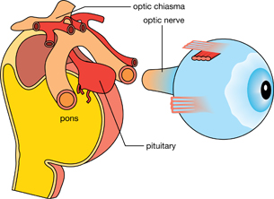
In FMEN1 specific endocrine glands, such as the parathyroid glands, pituitary gland, pancreas, and adrenal glands, or a combination of all, become overactive due to the growth of mostly benign (non-cancerous) tumours called neoplasms. These tumours disrupt normal function by crowding nearby structures and cells. For example, a tumour in the pituitary gland can press against surrounding pituitary tissue, thereby damaging the normal part of the tiny pituitary gland or the nerves that carry visual information from the eyes. In the diagram to the left, notice the very close arrangement of the pituitary gland and the optic nerve. This is one reason why Emily’s vision was blurred. In this module you will learn about the endocrine system from Emily’s perspective and experiences with FMEN1.
In this module you will explore the big question of how the body uses chemical messengers to respond to stimuli, including stress, to establish homeostasis.
In Module 2 you will explore the following focusing questions:
- How is the endocrine system organized, and how do its parts communicate with each other and other parts of the body?
- Who is the “boss” of the endocrine system, and how is control of the endocrine system managed?
- How do the major endocrine glands, including the anterior and posterior pituitary gland, the thyroid gland, the parathyroid glands, the pancreas, and the adrenal glands, contribute to homeostasis?
- What are the metabolic roles fulfilled by hormones in the maintenance of homeostasis, and what are the physiological consequences of hormonal imbalances?
- How do the nervous and the endocrine systems interact to maintain overall homeostasis?
In your journey through the endocrine system, you will find the answers to the focusing questions. These questions are also very similar to those that Emily and her parents asked her doctors. In particular, Emily and her family wanted to know what medical technologies could control the homeostatic imbalances so that Emily could try to lead as normal a life as possible.
To help you organize the concepts you learn in Module 2, and to provide you with a study aid for review before you complete the Module Assessment, you may choose to download the Concept Organizer for Module 2. Fill in this concept organizer with the ideas you master as you work through each lesson, or prepare the organizer when you have completed Module 2. You can use keywords, point form, or any amount of detail that meets your needs. You may choose to work from the file on your computer, print the document and work from the paper copy, or copy the outline onto a large sheet of poster paper. After you have prepared your concept organizer, you may wish to check your work with the concept organizer provided in the Module Summary. The concept organizer provided outlines some of the key topics that you should include in each lesson of your concept organizer. This is a great tool to review and use for study purposes, but using this organizer is completely your choice.
The Module Assessment for Module 2 will present you with a case study involving research data from two people, one of whom is Emily. Your assessment will be a written response based on analysis of the research data. You will be expected to integrate and explain concepts, describe processes, identify technological solutions, and illustrate your skill in drawing graphs and feedback loops. When you have completed the module, go to the Module Summary and Assessment page for details about the case study and an evaluation rubric.
1.2. In this Module
Module 2—The Endocrine System
In This Module
Inquiry Question: How does the endocrine system deal with stress?
There are six lessons in Module 2.
Most of the lessons are designed to take you 80 minutes to complete; however, some lessons may take longer because of the significance of the concept being covered in the lesson. The suggested lesson times do not include the time needed to complete such activities as “Try This,” “Watch and Listen,” assignments, practice questions, review, or research.
This module corresponds to “Chapter 13: Hormonal Regulation of Homeostasis” on pages 434 to 467 in your textbook. You may choose to briefly read through these pages for an overview before you begin this module.
Lesson 1—Structure and Organization of the Endocrine System
Your endocrine system responds to stress (any change in your internal or external environment) with glands releasing biochemical messengers called hormones. Hormones help maintain the body’s homeostasis. Emily’s body experiences stress, but her glands don’t secrete hormones appropriately to establish homeostasis. What causes this difference in hormonal communication?
In order to explore these concepts, you will investigate the following focusing question:
- How is the endocrine system organized, and how do its parts communicate with each other and with other various parts of the body?
Lesson 2—Who Is in Charge?
The endocrine system needs a master, or boss, to ensure homeostasis. This is the hypothalamus-pituitary gland complex.
In this lesson you will explore the following question:
- Who is the boss of the endocrine system, and how is control of the endocrine system managed?
Lesson 3—The Adrenal Gland
“Fight-or-flight” reaction! What would it be like going through life with a constant adrenaline rush?
In this lesson you will focus on the following question:
- How do the adrenal gland and its secretions affect the body?
Lesson 4—The Thyroid and Parathyroid Glands
If you have hyperthyroidism, you may always feel too hot, experience weight loss, find it difficult to concentrate, and be jittery and hyperactive. If you have hypothyroidism, you might always be cold, overweight, tired, and lethargic.
To understand the role of the thyroid gland in the functioning of the endocrine system, you will explore the following focusing question:
- How do the thyroid and parathyroid glands contribute to homeostasis?
Lesson 5—The Pancreas
Before lunch you are hungry, tired, and light-headed because of your low blood sugar level. What do you grab to eat? A candy bar or a tuna sandwich? Or do you just skip lunch? The pancreas manages blood sugar levels. For a diabetic, this is a daily balancing act—one that can have very serious consequences if it is not regulated.
To understand the role of the pancreas in the endocrine system, you will investigate the following focusing question:
- How does the pancreas contribute to homeostasis?
Lesson 6—Bringing It Together
The endocrine system responds to stimuli by using chemical messengers in the blood to control metabolic processes in the body. Your body is kept in balance because your nervous system responds to immediate stimuli or crises while your hormonal system maintains long-term stability.
This lesson will explore how the nervous and endocrine systems complement one another and communicate with each other to maintain homeostasis and ensure your well-being by investigating the following focusing question:
- How are the nervous and endocrine systems interdependent, and how are they different?
1.3. Lesson 1
Module 2—The Endocrine System
Lesson 1—Structure and Organization of the Endocrine System
 Get Focused

© mountainberryphoto/iStockphoto
In the Big Picture you were introduced to Emily and her struggle with familial multiple endocrine neoplasia type 1 (FMEN1) disorder. You might think that you experience stress when you are cramming for exams or are worried about a friend. You may get a headache, your heart may race, or you may have a sleepless night. Your endocrine system reacts to the stress by secreting many hormones that help you cope with the stress. When the exam or the crisis has passed, you seem to calm down because your endocrine system has returned the level of “stress” hormones to normal. In Emily’s case, her body experiences constant stress with no chance of relief. How might Emily’s endocrine system be different from yours? You both have the same organs, but it is obvious something is not working or communicating properly in Emily’s case.
In order to explore these concepts, you will investigate the following focusing question:
- How is the endocrine system organized, and how do its parts communicate with each other and with various parts of the body?
 Module 2: Lesson 1 Assignment
Your teacher-marked Module 2: Lesson 1 Assignment requires you to submit a response to the following:
- Lab—Student Stress
- Reflect and Connect
Download a copy of the Module 2: Lesson 1 Assignment to your computer now. You will receive further instructions on how to complete this assignment later in the lesson.
The other questions in this lesson are not marked by the teacher; however, you should still answer these questions. The Self-Check and Try This questions are placed in this lesson to help you review important information and build key concepts that may be applied in future lessons.
After a discussion with your teacher, you must decide what to do with the questions that are not part of your assignment. For example, you may decide to submit to your teacher the responses to Try This questions that are not marked. You should record the answers to all the questions in this lesson and place those answers in your course folder.
While you are completing this lesson, there will be many opportunities for you to acquire, understand, and practise the concepts that are presented to you. As you complete these activities, as well as your summary notes, you will file everything in your course folder to reference when you are preparing for exams.
Remember you also have the option of trying additional questions from the textbook for further practice. Consult with your teacher for the answers to these questions. The Key will also provide you with many Diploma Exam-style multiple-choice, numerical-response, and written-response questions that will be an excellent review of the module. Practising your responses to these types of questions is good preparation for the Diploma Exam.
1.4. Page 2
Module 2—The Endocrine System
 Explore
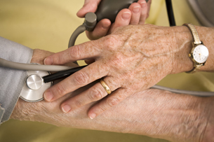
 Lab—Student Stress

© absolut/StockXpert
Think about how you feel when you are worried about exams, deadlines, sick friends, or a relationship. Your body responds with a racing heart beat, sweaty hands, fast breathing, and, maybe, sleeplessness.
In this activity you will measure one or, if possible, both of the following physiological changes:
- pulse, which is an indirect measure of heart rate
- blood pressure, which is the pressure exerted against blood vessel walls as circulating blood passes through the vessels
Note that measuring blood pressure will be dependent on the availability of a blood pressure monitor. You will complete the lab questions in the Lesson 1 Assignment. You will submit the data table, your responses to the four “Analysis” questions, and the conclusion to your teacher for assessment.
Problem (Purpose)
How does a stressful situation affect pulse and blood pressure?
Materials
- printed test questions provided by your teacher
- a stopwatch or a watch or clock with a second hand
- a blood pressure monitor: If you do not have a blood pressure monitor at home, contact your teacher to discuss your options. Many drugstores and pharmacies have blood pressure machines onsite. As well, the local health unit or a medical clinic may allow you to borrow a blood pressure monitor to complete this lab.
Procedure
Work with a partner.
Step 1: Using a watch with a second hand or a stopwatch, time your partner for 2 min as he or she sits in a comfortable chair with eyes closed and takes deep, relaxing breaths.
Step 2: When the 2-min “relaxation period” is up, take your partner’s pulse and measure his or her blood pressure. Look at the picture below for the correct placement of the fingers in order to feel the strongest pulse. If you are measuring the pulse manually, count the number of pulses in 15 s and then multiply by 4 to get the heart rate per minute. If you are using a digital blood pressure monitor, it may measure the pulse for you.
If you are using a digital blood pressure monitor, be sure to follow the instructions provided with the machine. If you are using a manual blood pressure cuff or a digital monitor, you can leave the deflated cuff on your partner’s arm for the next step.
Record the pulse and blood pressure in the data table in the Lesson 1 Assignment. These values will provide baseline data for your partner in a relaxed state.
© 2008 David Sucsy/istockphoto

© Franz Pfluegl 2006 - Fotostudio Pfluegl/istockphoto
Step 3: Note the time and administer the test to your partner. Your partner will have 2 min to complete the test questions provided by your teacher. Try to bother your partner by telling him or her to hurry up and that time is running out. This is your chance to stress out your partner!
Step 4: At the end of the 2 min shout, “Time is up!” Immediately take and record your partner’s pulse and blood pressure. Record the data for Reading 2 in the data table in the Lesson 1 Assignment. Check and record your partner’s pulse and blood pressure again after another 2 min (Reading 3), and then again 2 min after that (Reading 4).
Analysis
Complete the “Analysis” questions in the Lesson 1 Assignment.
Conclusion
In this lab, both the nervous and endocrine systems brought about the physiological changes that you measured. What conclusion can you make about how a stressful situation affects pulse and blood pressure? Which parts of the nervous and endocrine systems were involved? What can you conclude about the body’s ability to recover from stressful situations? How were the nervous and endocrine systems involved in this recovery? Write a response to these questions in the “Conclusion” part of the Lesson 1 Assignment.
1.5. Page 3
Module 2—The Endocrine System
Negative Feedback Loops
In the Student Stress lab you learned that a stressful situation, such as taking a test in a very short period of time, and being bothered while doing so, affects physiological processes. As a result, responses such as blood pressure and heart rate can change. After the stressful situation passes, heart rate and blood pressure return to normal or to “at-rest” values.
set point (set value): ideal or optimum conditions
negative feedback: a mechanism where deviation from the optimum state causes a return to the optimum state; acts to eliminate any deviation from optimal conditions and leads to stability
During every moment of your life, your body regulates carbon dioxide and oxygen levels, maintains water and salt balance, controls blood glucose levels, regulates temperature, maintains a regular heartbeat, and sustains a normal blood pressure. An organism is said to be in homeostasis when the internal environment is maintained at the balanced or best condition for each of these factors, which is called the set point. Homeostasis provides cells with a relatively constant environment, which helps them to work efficiently no matter what is going on outside the body.
Processes that aim to keep a potentially fluctuating value for a system within narrow limits use negative feedback mechanisms. Negative feedback means that when a condition changes, such as a drop in blood sugar level (BSL), the opposite effect is initiated, such as an effort to raise BSL. In a negative feedback system, there needs to be a sensor or receptor that measures the value of the feature to be controlled—for example, chemoreceptors monitor chemical levels in the body, such as the glucose concentration in the blood. If the sensor finds that the value is higher than it should be, it sends the information to an effector, which initiates a response to lower the value back toward the correct level. The effector continues this response until the sensor, which is still measuring the value, finds that the value is now too low. The receptor then sends information to the effector to stop doing whatever it is doing and start doing something to raise the value once more. Information is therefore “fed back” to the sensor from the effector. The feedback is called “negative” because it stops the effector from doing one thing and stimulates it to do the opposite. This is illustrated in the following diagram. Note: Although the term blood sugar level, BSL, is often used to describe the level of sugar in the blood, the term blood glucose level, or BGL, is technically more accurate.

In the case of regulation of blood glucose, an increase in the concentration of glucose in the blood sets into motion the processes that decrease the concentration. Conversely, a decrease in glucose concentration sets into motion the processes that increase the glucose concentration. The result is that the concentration of glucose in the blood automatically returns to its set value. This is shown in the diagram below.

 Read
Read
More information about homeostasis and feedback loops is found on page 203 of your textbook.
 Watch and Listen
Review the concept of negative feedback by watching the following segment of “The Hypothalamus and Pituitary: The Master Complex.” Select “Bio Fact: Negative Feedback System” from the menu.
You may be required to enter a username and password in order to access the video. Contact your teacher for this information.
Drawing a Feedback Loop
An increase in blood glucose can be represented by a feedback loop where the following conventions and symbols are used.
- An ascending arrow (↑) means an increase in the parameter.
- A descending arrow (↓) means a decrease in the parameter.
- A horizontal arrow (→) means “leads to.”
- A dashed horizontal arrow (→) with the words negative feedback or the negative symbol, sometimes in a circle, means that the opposite effect has been performed.

1.6. Page 4
Module 2—The Endocrine System
 Self-Check
SC 1. Now it is your turn to try your hand at drawing a feedback loop using all of the correct conventions and symbols described earlier. Given the following information about the hormone ADH (antidiuretic hormone) and its regulation of water volume in the body, summarize by drawing a negative feedback loop. After checking your answer, store your feedback loop in your course folder. Use it as an example when reviewing how to draw feedback loops. This is an important skill in Biology 30. Drawing diagrams, feedback loops, and flow charts is often part of the open-response essay question on the Diploma Exam. In Module 2 you will have an opportunity to read about, study, and draw many feedback loops.
Harjinder is losing water through perspiration while playing three-on-three basketball on a hot summer day.
- Harjinder forgot to bring a water bottle to the basketball court and has no water to drink.
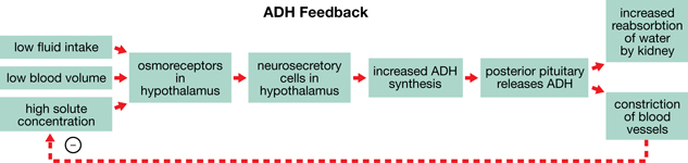
- This situation results in a decrease in the plasma (watery part) of the blood, so the solute (dissolved substances) concentration of the blood increases. As a result, both the blood volume and the blood pressure decrease.
- Sensors (or receptors) in the hypothalamus, called osmoreceptors, sense the increased solute concentration, decreased blood volume, and decreased blood pressure.
- The hypothalamus sends messages to the posterior pituitary gland to increase the release of the hormone ADH.
- An increased level of ADH causes the kidneys (kidney tubules) to retain (reabsorb) more water and release less in the urine.
- Reabsorbed water in the blood increases blood volume, increases blood pressure, and decreases solute concentration.
- Information about the preceding three factors is fed back to the sensors in the hypothalamus.
Self-Check Answers
SC 1. Your negative feedback loop should resemble the one shown below. It can be drawn horizontally; vertically, like the example shown on page 441 in your textbook; or in a combination of horizontal and vertical arrangements, like the diagram below. If you experienced difficulty drawing the negative feedback loop, or if you do not understand the concept, consult with your teacher.

Positive Feedback

© AKV/shutterstock
Sometimes the deviation from the set point or normal value is not corrected. Instead it leads to a further deviation. The result is a “runaway” situation in which a change triggers more change in the same direction. This is called positive feedback. At first sight, positive feedback would appear to be damaging, and even destructive. It may cause illness or the onset of a disorder.
Returning to the glucose example, if the pancreas were not able to decrease blood glucose after a meal because it could not produce insulin, the glucose level would remain high. You would feel hungry and perhaps eat a candy bar. As the glucose was absorbed from the stomach and intestines, even higher blood glucose levels would result. You might know this as the disorder called diabetes mellitus, Type 1. You will study diabetes in considerable detail in Lesson 5 of this module.
Using the same conventions and symbols that you learned for drawing negative feedback loops, a positive feedback loop for glucose regulation might look like the following:

In some cases, positive feedback is a good thing. In Unit B you will study the process of natural childbirth, which is dependent on positive feedback involving the hormone oxytocin.
Self-Check
SC 2. Using the information below, practise drawing a positive feedback loop to show how water regulation functions when the hormone ADH cannot be secreted. Use the symbols and conventions that you learned when drawing a negative feedback loop. After comparing your work with the suggested answer, save your work in your course folder.
- Randall is losing water through perspiration while playing three-on-three basketball on a hot summer day.
- Randall forgot to bring a water bottle to the basketball court and has no water to drink.
- This situation results in a decrease in the plasma (watery part) of the blood, so the solute (dissolved substances) concentration of the blood increases and both the blood volume and the blood pressure decrease.
- Sensors (or receptors) in the hypothalamus, called osmoreceptors, sense the increased solute concentration, decreased blood volume, and decreased blood pressure.
- The hypothalamus sends messages to the posterior pituitary gland to increase the release of the hormone ADH.
- A tiny tumour in the posterior pituitary prevents the release of ADH.
- The kidney cannot reabsorb more water, so it is released as an increased volume of urine.
- This information is fed back to the sensors in the hypothalamus.
Self-Check Answers
SC 2. In this example, positive feedback reinforces the loss of water in Randall’s body, not only from perspiring and drinking less, but also from excreting increased amounts of urine. Your positive feedback loop should look similar to the one below.

1.7. Page 5
Module 2—The Endocrine System
Fluctuation Around the Set Point

© GOH SIOK HIAN/shutterstock
Do you recall playing on the seesaw at the playground? Do you remember how hard it was for you and your friend at the other end to balance the seesaw evenly? Usually, one of you would be above and the other would be below the balance point.


© Tomasz Trojanowski/shutterstock
In humans, the sensors and receptors are specialized cells. Some of these cells are in the brain, but cells in other organs, such as the pancreas, also act as detectors for particular substances. Many different organs act as effectors. For example, the skin is an effector in temperature regulation, while the kidneys are effectors in the regulation of water content. Information passes from sensors to effectors either along nerves, as in the fight-or-flight regulation of epinephrine from the adrenal medulla, or via hormones in the blood, as in regulation of water content. The overall response is co-ordinated by some kind of control centre or boss. In the endocrine system, the control centre is the hypothalamus-pituitary complex.
 Self-Check
To ensure that you understand how negative feedback loops contribute to homeostasis and how positive feedback affects this balance, answer the following questions. At the Diploma-Exam level, your response must be complete and well-expressed using Biology 30 vocabulary. Check your answers and store your work in your course folder.
SC 3. Define homeostasis and explain how the endocrine system helps to maintain homeostasis in the body.
Use the following information describing how the temperature in your house is maintained to answer question SC 4.
|
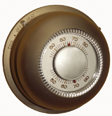
SC 4. Draw and describe a negative feedback mechanism for a thermostat connected to the furnace in your house. How is this feedback mechanism similar to how some hormones are regulated in the body?
Use the following information to answer question SC 5.
|
SC 5. Draw a positive feedback loop that results in a fever.
Self-Check Answers
SC 3. Homeostasis is the maintenance of relative constancy of the internal environment around an optimal value called the set point. The endocrine system is self-regulating and helps to regulate other body systems through negative feedback mechanisms.
SC 4. A negative feedback loop for a mechanical system, such as your heating system, might look like the following.

This mechanism of negative feedback is similar to how hormones regulate the internal environment.

1.8. Page 6
Module 2—The Endocrine System
Endocrine Glands

In Module 1 you learned about the basic unit of the nervous system—the neuron—and how communication occurs through neural pathways. Now, you will learn about the major glands of the endocrine system and how communication through the use of special chemical messengers called hormones occurs.
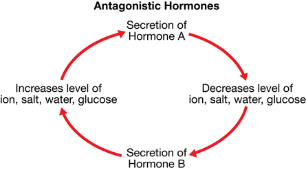
Endocrine glands are ductless glands—they do not release their secretions into a duct as exocrine glands do. Instead, endocrine glands secrete their hormones directly into the blood, which acts as the transport medium. As the hormones pass cells, only those cells with special receptors will react to their presence. These cells are called target cells. Lipid-soluble hormones and water-soluble hormones activate their target cells very differently. Hormones interact together and, in many instances, one hormone counteracts the action of a second hormone. Hormones such as these are called antagonistic hormones. This type of action is illustrated in the figure on the right.
Some hormones, called tropic hormones, influence other endocrine glands. They are secreted by the hypothalamus-pituitary complex. These hormones are very important in the control and regulation of the endocrine system. An important example is the gonadotropic hormones, which affect the reproductive organs. You can view the regulatory pathway of tropic hormones in “Figure 13.10” on page 441 of your textbook. You may want to sketch a copy this pathway and store it in your course folder for later reference.
hormone: a circulating chemical messenger that is produced by specialized cells, circulated in the bloodstream, and co-ordinates the various parts of the body by interacting with target cells
endocrine gland: a cell, tissue, or organ that produces secretions that are released directly into the bloodstream; a ductless gland; for example, the thyroid gland
exocrine gland: a cell, tissue, or organ that produces secretions that are moved through ducts or channels; for example, a sweat gland
target cells: cells with specialized receptor structures
When stimulated, these receptor structures cause a response in the target cells.
lipid-soluble hormone: a hormone that is chemically identified as a lipid or steroid, such as testosterone, estrogen, progesterone, or cortisol
water-soluble hormone: a hormone that is chemically identified as either an amino acid or a protein, such as epinephrine, human growth hormone, thyroxine, insulin, and glucagon
antagonistic hormones: two hormones that produce opposite effects
tropic hormone: a hormone that has another endocrine gland as its target cell
gonadotropic hormones: hormones that affect the reproductive organs; also called the gonads
 Read
Read
To help you identify the main endocrine glands, read pages 436 to 441 in your textbook. Summarize your readings about the endocrine glands in a chart or concept map with the following sections: “endocrine gland,” “hormone produced by gland,” and “primary effect of the hormone.” Place this chart in your course folder for future reference.
 Watch and Listen
To further explore and review these concepts visually, watch the following segments of “The Hypothalamus and Pituitary: The Master Complex”:
- “Endocrine System”
- “Hormones”
You may be required to enter a username and password to access these videos. Contact your teacher for this information.
 Self-Check
SC 6. Complete either Choice A or Choice B.
Choice A
This interactive drag-and-drop exercise has two parts.
In the first part of the exercise, match the statement that describes a hormone or the function of a hormone with the appropriate endocrine gland.
In the second part of the interactive exercise, drag the name of the hormone to the gland that produces it.
Choice B
Practise labelling the endocrine glands, identifying the hormones they produce, and describing the main functions of the hormones in the exercise that follows. After you have completed the exercise and checked your answers, file it in your course folder for access when you are studying.
Download the diagram of the endocrine system.
- Label the major endocrine glands numbered 1 through 8 on the diagram.
- List the hormones (the number of hormones to be identified is indicated in brackets) that each gland produces. The ovary and testis will be studied in Unit B. Although the thymus and pineal gland are part of the endocrine system, you are not responsible for studying them for this course.
- Construct and complete a table with the following headings.
Endocrine Gland |
Hormone |
Target Cells |
Primary Function |
 Self-Check Answers
Choice B
1. and 2. Your labels of the glands on the diagram and the hormones that the glands produce should be very similar to the ones that follow.
1. hypothalamus: secretes releasing and inhibiting factors/hormones
2a. anterior pituitary gland: secretes human growth hormone; thyroid stimulating hormone (TSH); adrenocorticotropic hormone (ACTH); and follicle stimulating hormone (FSH), luteinizing hormone (LH), and prolactin (The last three hormones will be studied in detail in Unit B.)
2b. posterior pituitary: releases oxytocin (to be studied in detail in Unit B) and antidiuretic hormone (ADH)
3. parathyroid glands: secrete parathormone (PTH)
4. thyroid gland: secretes thyroxine and calcitonin
5. adrenal glands; adrenal cortex secretes cortisol, aldosterone, and gonadotropins (to be studied in Unit B); adrenal medulla secretes epinephrine and norepinephrine
6. islet cells of pancreas; insulin and glucagon
7. ovary secretes estrogen and progesterone (to be studied in Unit B)
8. testis secretes testosterone and inhibin (to be studied in Unit B)
3. Your completed table should look like the following sample.
Endocrine Glands, Their Hormones, Target Cells, and Main Functions
Endocrine Gland |
Hormone |
Target Cells |
Primary Functions |
Hypothalamus |
releasing/inhibiting hormones |
pituitary gland |
regulates secretion by anterior pituitary |
Posterior Pituitary |
antidiuretic hormone (ADH) |
kidney |
increases water reabsorption |
oxytocin |
uterus/breasts |
stimulates contraction of uterus and release of milk by breast glands |
|
Anterior Pituitary |
thyroid stimulating hormone (TSH) |
thyroid gland |
stimulates thyroid gland to secrete thyroxine |
adrenocorticotropic hormone (ACTH) |
adrenal cortex |
stimulates adrenal cortex to secrete cortisol |
|
growth hormone (hGH) |
most cells |
stimulates cell division, growth of bones, and metabolism |
|
follicle stimulating hormone (FSH) |
ovary/testis |
stimulates production of egg and sperm cells |
|
luteinizing hormone (LH) |
ovary/testis |
stimulates production of male and female sex hormones |
|
prolactin |
breast cells |
stimulates milk production |
|
Adrenal Medulla |
epinephrine/norepinephrine |
most cells |
fight-or-flight hormones |
Adrenal Cortex |
cortisol |
most cells |
raises blood glucose and breaks down protein |
aldosterone |
kidney |
increases sodium retention and increases water reabsorption |
|
Parathyroid Glands |
parathormone (PTH) |
bone cells |
stimulates calcium release from bones |
Thyroid Gland |
thyroxine |
most cells |
increases metabolic rate and regulates growth and development |
calcitonin |
bone cells |
stimulates bones to absorb calcium |
|
Pancreas |
insulin |
most cells |
increases permeability of glucose in cells; stimulates liver cells to convert glucose to glycogen |
glucagon |
liver cells |
stimulates release of glucose by converting glycogen to glucose |
|
Ovary |
estrogen |
uterus/other cells |
stimulates growth of uterine lining; stimulates development of female secondary sex characteristics |
progesterone |
uterus |
promotes growth of uterine lining; inhibits uterine contractions during pregnancy |
|
Testes |
testosterone |
testis/other cells |
promotes sperm production; stimulates development of male secondary sex characteristics |
inhibin |
testis |
inhibits sperm production |
1.9. Page 7
Module 2—The Endocrine System
 Self-Check
To refresh your memory of the structure and organization of the endocrine system and the hormones that the endocrine glands produce, complete the following questions. Check your work. If you have any questions, discuss them with your teacher before you move on to the rest of this lesson.
SC 7. Maintaining stable conditions in the internal environment of a cell is called
- positive feedback
- homeostasis
- reflex loops
- tropic control
SC 8. Your body temperature is controlled by
- negative feedback
- positive feedback
- tropic hormones
- antagonistic hormones
SC 9. The three components of a homeostatic mechanism in the body are
- a stimulus, a sensor, and a response
- a receptor, a neural pathway, and a control centre
- a sensor, a set point, and a response
- a sensor, an effector, and a control centre
SC 10. Skeletal growth is promoted by a hormone secreted by the
- adrenal cortex
- adrenal medulla
- anterior pituitary
- posterior pituitary
SC 11. A hormone that regulates glucose levels in the blood and a hormone that regulates sodium ions in the blood, and indirectly regulates water reabsorption by the kidneys, are, respectively,
- aldosterone and insulin
- glucagon and aldosterone
- insulin and antidiuretic hormone
- epinephrine and antidiuretic hormone
SC 12. During an emergency situation, the adrenal gland is stimulated to release a hormone that directly causes an increase in
- conversion of glucose to glycogen
- insulin levels
- thyroxine levels
- blood glucose levels
Use the following information to answer question SC 13.
| A tumour of the adrenal medulla is called phenochromocytoma. This tumour causes hypersecretion of epinephrine and norepinephrine, and a number of other symptoms. |
SC 13. Possible symptoms of phenochromocytoma include
- increased heart rate, increased blood glucose, increased metabolic rate
- decreased heart rate, increased blood glucose, increased metabolic rate
- increased heart rate, decreased blood glucose, decreased metabolic rate
- decreased heart rate, decreased blood glucose, decreased metabolic rate
SC 14. The release of milk from the breast after the birth of a baby is related to increased levels of
- estrogen
- progesterone
- oxytocin
- FSH and LH
True-or-False Questions
Identify each of the statements below as being true or false. If you think that the statement is false, change the statement to make it true.
SC 15. The main purpose of the endocrine system is for the protection of the body.
SC 16. Hormones are released into the bloodstream.
SC 17. Adrenocorticotropic hormone is released by the adrenal glands to control the pituitary gland.
SC 18. Positive feedback leads to instability of the internal environment.
SC 19. The anterior pituitary releases tropic hormones that were produced by the hypothalamus and stored.
Self-Check Answers
SC 7. B
SC 8. A
SC 9. D
SC 10. C
SC 11. B
SC 12. D
SC 13. A
SC 14. C
SC 15. False
The main purpose of the endocrine system is maintaining homeostasis (a stable internal environment).
SC 16. True
SC 17. False
Adrenocorticotropic hormone is released by the anterior pituitary to control the adrenal cortex (adrenal gland).
SC 18. True
SC 19. False
You may have either of the following answers:
- The anterior pituitary synthesizes and releases tropic hormones.
- The posterior pituitary releases stored hormones that were produced in the hypothalamus.
1.10. Page 8
Module 2—The Endocrine System
 Reflect and Connect
You now have the opportunity to reflect on the structures and organization of the endocrine system by answering ten multiple-choice questions. Before you continue, glance through pages 436 to 442 and answer the questions on page 442 of your textbook. You can get feedback from your teacher about your responses to these questions.
 Module 2: Lesson 1 Assignment
Retrieve the copy of the Module 2: Lesson 1 Assignment that you saved to your computer earlier in this lesson. Complete the Reflect and Connect questions. Save your completed assignment in your course folder. You will receive instructions about when to submit your assignment to your teacher later in this lesson.
Reflect on the Big Picture

© Alexander Raths/shutterstock
At the beginning of this module, you were introduced to Emily. She has all the same endocrine glands as you do and she secretes all the hormones that you do; however, due to growth of tiny tumours in her endocrine glands, she may secrete too much of one kind of hormone and not enough of another. Her body is under constant stress, and her negative feedback mechanisms do not restore homeostasis. Instead, positive feedback results, and she becomes ill due to the oversecretion of hormones putting constant stress on her body.
 Going Beyond
Melatonin is a hormone produced by the tiny pineal gland in the brain. It is involved in your sleep-and-wake cycle. Melatonin is secreted during periods of darkness, causing you to sleep. Secretion is reduced during periods of light, causing you to be awake. Many people experience tiredness, inability to concentrate, and irritability when they are not exposed to sufficient periods of sunshine, such as during Canadian winters! Read “Light up Your Life!” on page 443 of your textbook and answer questions 1 and 2.
Module 2: Lesson 1 Assignment
Submit your completed Module 2: Lesson 1 Assignment to your teacher for assessment.
1.11. Page 9
Module 2—The Endocrine System
 Lesson Summary
In this lesson you investigated the following focusing question:
- How is the endocrine system organized, and how do its parts communicate with each other and with various parts of the body?
To answer this question, the major glands of the endocrine system, including the hypothalamus-pituitary complex, the thyroid gland, the parathyroid glands, the adrenal glands, and the islet cells of the pancreas, were introduced and studied. These glands secrete hormones, which have unique effects on the body by interacting with target cells. Some of these hormones work opposite each other and are called antagonistic hormones. Other hormones, called tropic hormones, target other endocrine glands, causing them to secrete more hormones. Levels of hormonal secretions are regulated by negative feedback mechanisms. Negative feedback tends to stabilize a system because the response compensates for the change in the internal environment. This leads to the re-establishment of homeostasis. Reinforcing the change in the internal environment leads to instability and an imbalance or deviation from homeostasis called positive feedback. It is rarely beneficial in the body.
As a review of the concepts of this lesson, you may wish to view all of “The Hypothalamus and Pituitary: The Master Complex.”
You may be required to enter a username and password to access this video. Contact your teacher for this information.
Lesson Glossary
Consult the glossary in the textbook for other definitions that you may need to complete your work.
antagonistic hormones: two hormones that produce opposite effects
endocrine gland: a cell, tissue, or organ that produces secretions that are released directly into the bloodstream; a ductless gland; for example, the thyroid gland
exocrine gland: a cell, tissue, or organ that produces secretions that are released through ducts or channels; for example, a sweat gland
gonadotropic hormones: hormones that affect the reproductive organs; also called the gonads
hormone: a circulating chemical messenger that is produced by specialized cells, circulated in the bloodstream, and co-ordinates the various parts of the body by interacting with target cells
lipid-soluble hormone: a hormone that is chemically identified as a lipid or steroid, such as testosterone, estrogen, progesterone, or cortisol
negative feedback: a mechanism where deviation from the optimum state causes a return to the optimum state; acts to eliminate any deviation from optimal conditions and leads to stability
positive feedback: a mechanism where movement away from the optimum state causes further deviation from the optimum state; usually leads to instability and is tolerated by the body only for a short time
set point (set value): ideal or optimum conditions
target cells: cells with specialized receptor structures
When stimulated, these receptor structures cause a response in the target cells.
tropic hormone: a hormone that has another endocrine gland as its target cell
water-soluble hormone: a hormone that is chemically identified as being either an amino acid or a protein, such as epinephrine, human growth hormone, thyroxine, insulin, and glucagon
1.12. Lesson 2
Module 2—The Endocrine System
Lesson 2—Who Is in Charge?
 Get Focused

In the first lesson of this module, you learned about the different glands that make up the endocrine system. The level of hormones circulating in the body from these different glands needs to be just right. Like the brain in the nervous system, the endocrine system also has a "boss" to ensure homeostasis—the hypothalamus-pituitary gland complex. Module 1 presented the hypothalamus as part of the brain, making this structure one of the places where the nervous and endocrine systems overlap. You may know the pituitary as the gland that regulates cellular respiration or metabolism from your studies in Biology 20. If this complex were not doing its job, your life would quickly unravel. You would not be able to function or develop normally. Your growth might be stunted and, if you were a boy, your voice would never change. In Emily’s case, tiny tumours in her pituitary gland cause either an oversecretion or undersecretion of several pituitary hormones that regulate other endocrine glands. Because they are inappropriately stimulated, these glands, in turn, oversecrete or undersecrete their hormones and cause potentially life-threatening symptoms, such as imbalances in blood sugar level.
In this lesson you will explore the following focusing question:
- Who is the boss of the endocrine system, and how is the control of the endocrine system managed?
 Module 2: Lesson 2 Assignment
Your teacher-marked Module 2: Lesson 2 Assignment requires you to submit a response to the following:
- Thought Lab—Evaluating Potential Uses for Human Growth Hormone
Download a copy of the Module 2: Lesson 2 Assignment to your computer now. You will receive further instructions on how to complete this assignment later in the lesson.
You must decide what to do with the questions that are not marked by the teacher.
Remember that these questions provide you with the practice and feedback that you need to successfully complete this course. You should respond to all the questions and place those answers in your course folder.
While you are completing this lesson, there will be many opportunities for you to acquire, understand, and practise the concepts that are presented to you. As you complete these activities, as well as your summary notes, you will file everything in your course folder to reference when you are preparing for exams.
Remember you also have the option of trying additional questions from the textbook for further practice. Consult with your teacher for the answers to these questions. The Key will also provide you with many Diploma Exam-style multiple-choice, numerical-response, and written-response questions that will be an excellent review of the module. Practising your responses to these types of questions is good preparation for the Diploma Exam.
1.13. Page 2
Module 2—The Endocrine System
 Explore
The Posterior Pituitary
The hypothalamus lies just above the tiny, pea-sized pituitary gland in the middle of the head. The two structures are connected by a stalk. Anatomically, the pituitary gland is made up of two very different kinds of cells. The posterior pituitary is made up of modified nerve cells. It is connected to the hypothalamus by neurosecretory cells in the stalk that secrete two hormones—antidiuretic hormone (ADH, also called vasopressin) and oxytocin. Releasing hormones (releasing factors) from the hypothalamus signal the release of the stored ADH and oxytocin from the posterior pituitary into the bloodstream.
In Biology 20 you learned that ADH targets cells in the kidney to reabsorb more water into the blood, producing smaller volumes of concentrated urine. In Lesson 1 you practised drawing the negative feedback loop that regulates ADH production. If ADH cannot be produced or secreted, copious amounts of urine are produced (up to 24 L in 24 h). The person experiences what is known as diabetes insipidus (from diabetes, which means "to pass through," and insipidus, which means "without taste"), which is treated with ADH in pill form.
hypothalamus: the region of the brain located below the cerebral hemispheres and thalamus and just above the pituitary gland; functions in maintaining homeostasis, and is especially important in co-ordinating the endocrine and nervous systems; secretes hormones of the posterior pituitary as well as releases hormones that regulate the anterior pituitary
posterior pituitary: an extension of the hypothalamus composed of nervous tissue that secretes hormones produced in the hypothalamus into the bloodstream; consists of a temporary storage site for hormones produced in the hypothalamus
neurosecretory cells: specialized nerve cells in the hypothalamus that extend into the posterior pituitary and secrete ADH and oxytocin into the posterior pituitary and, subsequently, into the bloodstream
vasopressin: also called antidiuretic hormone (ADH)
diabetes insipidus: a condition caused by a lack of ADH, which results in excessive production of very dilute urine
ADH may be produced in insufficient quantities by the hypothalamus or the posterior pituitary may fail to release it into the bloodstream when a tumour develops.

© webphotographeer/iStockphoto
Oxytocin, the other hormone secreted by the hypothalamus and released by the posterior pituitary, begins the contractions of the uterine muscles at the end of pregnancy, thus initiating childbirth. It also causes tiny cells that surround the milk glands in the breasts to contract and squeeze milk into the nipples. You will study oxytocin in more detail in Unit B.
 Read
Read
To understand how ADH and oxytocin are part of the endocrine system, and to prepare for the other hormones you will study in this module, read from the top of page 444 to the heading “The Thyroid Gland: A Metabolic Thermostat” on page 446 in your textbook. You may wish to start a concept map similar to the chart on page 445 to facilitate your study of human hormones.
 Watch and Listen
To review and further your understanding of the hormones associated with the posterior pituitary, watch the following segments of “The Hypothalamus and Pituitary Gland: The Master Complex.” You may be required to enter a username and password to access these videos. Contact your teacher for this information.
- “Oxytocin”
- “Antidiuretic Hormone (ADH)”
- “Action of ADH”
- “Bio Fact: Altered Levels of ADH”
 Self-Check
To further your understanding and application of concepts related to posterior pituitary hormones, complete the following questions. Answer in full sentences where appropriate. Save your work in your course folder.
SC 1. In general, how is a hormone able to recognize and stimulate its target cells?
SC 2. Describe how the secretion of ADH is regulated by negative feedback. Include a feedback loop in your answer.
SC 3. What causes diabetes insipidus? Describe the symptoms of this condition.
SC 4. Copy and complete the following table.
Hormone |
Produced By |
Released By |
Function(s) |
Oxytocin |
|||
ADH |
Self-Check Answers
SC 1. A hormone is able to recognize its target cells because the target cells have receptors on their cell membranes that have a complementary shape to the shape of the hormone molecules. When the hormone fits into the receptors, the cells are stimulated by the hormone.
SC 2. When the solute concentration or osmotic concentration of the blood increases, tiny receptors called osmoreceptors in the hypothalamus are stimulated. In turn, neurosecretory cells in the hypothalamus are stimulated to secrete ADH, which moves along the axons of the neurosecretory cells into the posterior pituitary and is released into the bloodstream. ADH moves in the blood to the kidneys, where it fits into receptors on the kidney tubules. This causes the tubules to become more permeable and reabsorb more water into the blood, which, in turn, reduces the urine volume. The increased water in the blood reduces the solute concentration or osmotic concentration, which inhibits the osmoreceptors, and less ADH is released. Your diagram of a feedback loop should be similar to the one shown below.
SC 3. Diabetes insipidus is usually caused by a tumour that prevents the hypothalamus from producing ADH or prevents the posterior pituitary from releasing the ADH. Consequently, the kidney tubules cannot reabsorb adequate amounts of water, which, in turn, increases the urine output. The main symptom of diabetes insipidus is a very large output of urine, with the other components of urine remaining stable.
SC 4. Your completed table should resemble the following sample response.
Hormone |
Produced By |
Released By |
Function(s) |
Oxytocin |
hypothalamus |
posterior pituitary |
initiates uterine contractions at the end of pregnancy, thus starting parturition
initiates release of milk from the breast |
ADH |
hypothalamus |
posterior pituitary |
stimulates kidney tubules to reabsorb water into blood, which increases blood volume
stimulates constriction of blood vessels, thereby raising blood pressure |
1.14. Page 3
Module 2—The Endocrine System
Anterior Pituitary
anterior pituitary: an endocrine gland consisting of secretory cells that synthesize and secrete several hormones directly into the bloodstream
Compared to the posterior pituitary, which only stores and releases hormones produced in the hypothalamus, the anterior pituitary synthesizes and releases six major hormones. It is connected to the hypothalamus by blood vessels that run through the stalk. Three of the hormones (FSH, LH, and prolactin) are involved in the reproductive process, and will be studied in detail in Unit B. In this unit you will learn their general functions. The other three hormones, adrenocorticotropic hormone (ACTH), thyroid stimulating hormone (TSH), and human growth hormone (hGH), will be introduced in this lesson.
The hypothalamus and pituitary control the production and release of these hormones through negative feedback loops. The hypothalamus is stimulated to secrete releasing hormones by conditions in the internal environment of the body. Moving through the blood vessels in the stalk, the releasing hormones stimulate target cells in the anterior pituitary, which produces ACTH, TSH, and hGH. As indicated by its name, ACTH stimulates the outside layer of the adrenal gland, called the adrenal cortex, which releases cortisol, a major stress hormone, and aldosterone. TSH stimulates the thyroid gland and its production of thyroxine.
Emily’s anterior pituitary gland overproduces ACTH and undersecretes TSH. You will study the effects of inappropriate amounts of these hormones on the body in the next two lessons.
 Read
Read
releasing hormones: hormones produced by neurosecretory cells in the hypothalamus that stimulate or inhibit the secretion of hormones by the anterior pituitary; sometimes called releasing factors
Releasing hormones, which are produced by the hypothalamus, and ACTH, TSH, and hGH, which are produced by the anterior pituitary, are called tropic hormones. In Lesson 1 you learned that tropic hormones are one group of hormones. Do you remember their characteristics? Explore this group of hormones in more depth by studying “Figure 13.10” on page 441 of your textbook. Read and summarize the section about tropic hormones, “Regulating the Regulators,” on pages 441 and 442 of your textook. Store your notes in your course folder.
 Watch and Listen
You can review some of the concepts on the anterior pituitary by watching the following segment of “The Hypothalamus and Pituitary Gland: The Master Complex.” You may be required to enter a username and password to access this video. Contact your teacher for this information.
- “Anterior Pituitary Gland”
 Self-Check
SC 5. Practice matching the structures of the hypothalamus-pituitary complex with their hormones by completing a drag-and-drop activity.
To practise applying your knowledge about specific tropic hormones and their regulation by the hypothalamus and anterior pituitary gland, complete the following questions.
SC 6. Using ACTH as an example, explain a tropic hormone. Besides ACTH, identify three other tropic hormones.
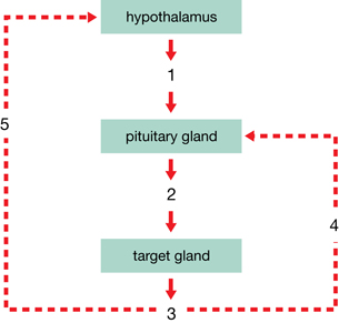
Use the following flow chart, which illustrates regulation by tropic hormones, to answer questions SC 7 through SC 12.
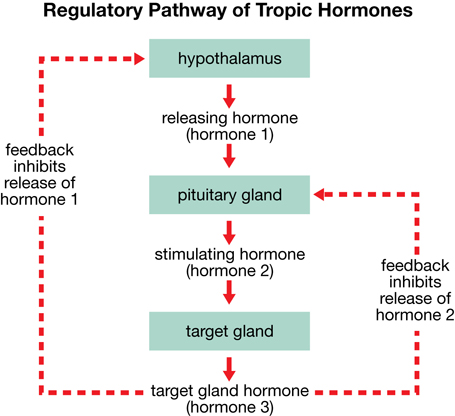
SC 7. How is the secretion of tropic hormones from the pituitary gland regulated?
SC 8. If hormone 2 in the tropic hormone pathway is TSH, what is the target gland?
SC 9. If hormone 2 is TSH, what is hormone 3?
SC 10. Explain how hormone 3 can inhibit or stimulate the release of hormone 1 and inhibit release of hormone 2?
SC 11. If the target gland is the adrenal cortex, identify the specific tropic hormone at work.
SC 12. Identify hormone 3 produced by the adrenal cortex.
SC 13. Explain how the anterior and posterior pituitaries differ with respect to their relationship to the hypothalamus.
SC 14. Explain how the differences between the two regions of the pituitary relate to the nature of their hormonal secretions.
SC 15. Suppose a scientist has discovered a new hormone. It is not clear what gland produces the hormone, but people who produce above-average amounts of this hormone also produce very high levels of insulin. Based on your knowledge of how tropic hormones function, provide a possible explanation for the observation.
Self-Check Answers
SC 6. ACTH is an example of a tropic hormone because it is released by the anterior pituitary and travels through the blood to the adrenal cortex of the adrenal gland (another endocrine gland), which it stimulates to release hormones such as cortisol.
Other tropic hormones include releasing hormones from the hypothalamus, thyroid stimulating hormone (TSH), human growth hormone (hGH), LH, and FSH.
SC 7. The secretion of tropic hormones from the pituitary gland is regulated by releasing hormones from the hypothalamus, which either stimulate or inhibit the anterior pituitary gland.
SC 8. If hormone 2 in the tropic hormone pathway is TSH, the target gland is the thyroid.
SC 9. If hormone 2 is TSH, hormone 3 is thyroxine.
SC 10. Excessive amounts of thyroxine (hormone 3) inhibit the anterior pituitary gland from releasing TSH and the hypothalamus from secreting releasing hormones. The thyroid gland is inhibited and does not release thyroxine, thus lowering the amount circulating in the blood. Inadequate amounts of thyroxine stimulate the hypothalamus to secrete releasing hormones, which stimulates the anterior pituitary to release TSH, which stimulates the thyroid gland to release more thyroxine.
SC 11. If the target gland is the adrenal cortex, then the tropic hormone is ACTH.
SC 12. The adrenal cortex is stimulated to produce hormones such as cortisol.
SC 13. The posterior pituitary is an extension of the hypothalamus and is composed of specialized neurons, while the anterior pituitary is a true endocrine gland and is composed of several types of secretory cells that produce and release hormones. Both lobes of the pituitary gland are connected to the hypothalamus by a stalk, but the posterior pituitary is connected to the hypothalamus by neurons, while the anterior pituitary is connected to the hypothalamus by blood vessels.
SC 14. The neurosecretory cells of the hypothalamus secrete the hormones and move them along the axons to the posterior pituitary, where they are stored and released into the bloodstream as needed. Neurons from the hypothalamus stimulate the secretory cells of the anterior pituitary to synthesize the hormones, and releasing hormones circulating in the bloodstream stimulate the release of the anterior pituitary hormones.
SC 15. Since tropic hormones stimulate another gland to produce a hormone, the above-average amount of this new hormone could be what is stimulating the pancreas to secrete high levels of insulin. This new hormone might be produced by the hypothalamus or perhaps by the anterior pituitary.
1.15. Page 4
Module 2—The Endocrine System
© Sandra G/shutterstock
Human Growth Hormone
The anterior pituitary regulates growth, muscle development, and fat metabolism through the production of human growth hormone (hGH), which ultimately affects every part of your body. Some athletes are interested in using human growth hormone to improve performance because it stimulates protein synthesis and subsequent muscle development as well as toning of muscles. Some overweight people are interested in using this hormone to fight obesity because it inhibits the storage of fat and encourages its use for cellular respiration. At the same time, hGH inhibits use of carbohydrates such as glucose. Some older people are also interested in using hGH. As one ages, the levels of this hormone decrease—fat is harder to keep off and muscles become flabby. Testimonials by aging stars, such as Sylvester Stallone, are prominent in advertisements for hGH. You will examine some of these ideas in “Thought Lab: Evaluating Potential Uses for Human Growth Hormone.”
© Tiplyashin Anatoly/shutterstock
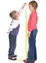
Nearly everyone is interested being tall. Fairytales often contain stories of maidens being saved by “tall, dark, and handsome” heroes. Human growth hormone stimulates the growth plates at the end of the long bones and causes these bones to lengthen and increase a person’s height. Too much hGH during childhood causes gigantism. Today (early 2009), Leonid Stadnyk is the tallest living person, at a height of 2.57 m or 8 feet 5.5 inches. Too little hGH during childhood results in pituitary dwarfism. Thapa Magar, the shortest living person, is only 50.8 cm or 20 inches tall.
Emily, who you met earlier, had some very small tumours develop in the anterior pituitary several years after her final growth spurt. Instead of gigantism, she shows the symptoms of acromegaly. Because her growth plates have sealed, she cannot grow in height. Instead, her jaw has thickened, as have her ribs; and her fingers, toes, and nose have become enlarged. This type of growth results from excessive production of hGH during adulthood.
gigantism: a condition where a person produces excess hGH during childhood, resulting in a height of 8 feet or more
pituitary dwarfism: a condition where a person does not produce enough hGH during childhood, resulting in short stature
acromegaly: a condition brought about by excessive secretion of human growth hormone in an adult; hands, feet, and nose widen and enlarge; jaw protrudes; voice becomes husky; barrel chest may develop; sweat glands enlarge; heart enlarges; high blood pressure may develop; enlarged tissues press on nerves, especially optic nerves, causing loss of vision, particularly in the outer fields; and pressure on the brain causes severe headaches
There is an increased likelihood of developing diabetes mellitus.
 Read
Read
To learn more about human growth hormone, and to summarize the functions and effects of this important hormone, you may wish to review pages 444 to 446 in your textbook. “Figure 13.11,” on page 444, and “Figure 13.15,” on page 446, are good summaries that you may want to copy and include in your course folder.
1.16. Page 5
Module 2—The Endocrine System
 Watch and Listen
To explore some interesting concepts about human growth hormone, watch the following segments of “The Hypothalamus and Pituitary Gland: The Master Complex,” and make some additional notes for your course folder. You may need to enter a username and password to access these videos. Contact your teacher for this information.
- “Human Growth Hormone”
- “Bio Challenge: Growth Hormone Imbalances”
- “Bio Discovery: Growth Hormone Deficiency”
- “Bio Review: Growth Hormone Therapy”
- “Bio STS: Synthetic Human Growth Hormone”
- “Bio Bit: Bovine Growth Hormone
 Try This
TR 1. Emily’s husband has become very concerned about some of the symptoms she is currently exhibiting, and the inability of several physicians to diagnose and treat whatever is wrong. One doctor figured out what was wrong, but he could only provide Emily and her husband with limited information. Emily’s husband has decided to write to a newspaper columnist who specializes in answering questions about health.
Pretend that you are Dr. Stoppain, who answers health-related questions in a daily newspaper column. You received the following letter, and your task is to write a response by defining the health condition, explaining the symptoms of the condition, outlining possible treatment, and answering the writer’s questions.
Dear Dr. Stoppain,
Concerned Husband |
Try This Answers
The following is a possible reply from Dr. Stoppain. Check your response to see whether you have outlined the cause of acromegaly, described at least two symptoms of the disorder, explained the ones that the husband mentioned in his letter (violent headaches, loss of peripheral vision), and outlined how acromegaly may be treated.
Dear Concerned Husband,
Acromegaly is a rare condition that is linked to a tumour of the pituitary gland, which causes the anterior pituitary gland to produce too much human growth hormone. The pituitary gland lies on the underside of the brain at a level approximately in line with the bridge of the nose.
In children, human growth hormone allows for the attainment of height programmed by the genes. The bones lengthen at growth plates on their ends. At the end of our growing period, these plates seal and no more growth is possible. In adults, too much growth hormone leads to radical changes in body appearance, but the changes occur so slowly that they often go unrecognized. Hands and feet become thick and wide. The forehead enlarges and the jaw juts out. Spaces appear between the teeth. Blood pressure rises and the heart enlarges.
Headaches are a common sign of a pituitary tumour. Vision problems also arise because the pituitary gland lies behind the optic nerve, and a pituitary tumour presses on the nerve and interferes with transmission of visual images to the brain.
Diagnosis of acromegaly is established by measuring growth hormone levels in the blood. A scan of the brain can clearly detect the presence of a tumour.
Treatment consists of removal of the tumour, which can often be done with instruments passed through the nose or palate. When surgery is not feasible, medicines can dampen the production of growth hormone. Sometimes, focused radiation therapy can shrink the tumour.
Your wife might not have any noticeable physical changes associated with acromegaly. Now that the condition has been proven, she can undergo appropriate therapy. Her life will not be shortened.
Dr. Stoppain |
 Going Beyond
Have you ever considered the problems that people of short stature encounter? If you have an opportunity to watch a rerun of a documentary called Short and Male, you should do so. It originally aired on CTV on Saturday, May 24, 2008. Thought-provoking questions are raised and the risks and benefits of hGH therapy are addressed. You might find it on the Internet by using search words such as “CTV,” “W5,” or “short and male.”
1.17. Page 6
Module 2—The Endocrine System
 Thought Lab—Evaluating Potential Uses for Human Growth Hormone
You will complete this Thought Lab in the Module 2: Lesson 2 Assignment.
Recent studies have linked job success with being tall, and many jobs have specifice physical requirements, including those for height and weight. Since 1985, genetic-engineering technology has been used to produce human growth hormone, referred to as synthetic human growth hormone. Some parents have pestered doctors to prescribe it for the purpose of increasing the stature of their children and, therefore, their potential for success. To add to the appeal of synthetic HGH, it has been discovered that human growth hormone may have some anti-aging properties. The Internet is littered with ads for its purchase.
Since the approval of the restricted use of synthetic hGH, concerns have arisen about its use and potential abuse. Health Canada has approved extremely limited use of the hormone, which is very expensive (injections cost in excess of $25 000 per year) and may be associated with several negative health effects.
Problem
Should Health Canada approve the widespread use of synthetic hGH for Canadians?

© Konstantin Sutyagin/shutterstock
Issue 1
Until recently, the use of synthetic hGH was approved only for those children who had malfunctioning pituitary glands and could not produce adequate amounts of the necessary hormone themselves. Recently, the use of synthetic hGH has been approved for children who are genetically of short stature. Should people have the option to take synthetic hGH just to increase their genetically predetermined height?
Issue 2

© Glenda M. Powers/shutterstock

© Carlos E. Santa Maria/shutterstock
Issue 3
Because one of the functions of hGH in the body is to build lean muscle mass, its use has become widespread among some athletes in various sports. In fact, many athletes at the 1996 summer Olympic Games in Atlanta, Georgia, referred to the event as the “hGH Games.” Despite its expense, many athletes, from baseball players to weightlifters, are acquiring synthetic hGH because it is difficult for drug testers to detect. Should competitive athletes be allowed legal access to synthetic hGH?
 Module 2: Lesson 2 Assignment
Retrieve the copy of the Module 2: Lesson 2 Assignment that you saved to your computer earlier in this lesson. Complete all of the questions. Save your completed assignment in your course folder. You will receive instructions about when to submit your assignment to your teacher later in this lesson.
1.18. Page 7
Module 2—The Endocrine System
 Reflect and Connect
So, who is in charge of the endocrine system? Is it the hypothalamus, the pituitary gland, or both? To review the connections between the hypothalamus and the pituitary gland, watch the following segment of “The Hypothalamus and Pituitary: The Master Complex.” You may need to enter a username and password to access this video. Contact your teacher for this information.
- “Relationship Between the Hypothalamus and the Pituitary Gland”
Then, using what you have learned in this lesson about the hypothalamus, the posterior pituitary, and the anterior pituitary, decide which you think is the boss of the endocrine system. Provide as much evidence as possible to support your position. You may choose the hypothalamus, the posterior pituitary, anterior pituitary, or a combination of these structures. You may wish to discuss your ideas with your teacher or post your position and evidence on the discussion board for your peers to evaluate. Read at least two other students’ positions and discuss their evidence.
 Self-Check
Complete the following multiple-choice questions, which will help assess your understanding of the concepts presented in this lesson. Discuss any questions that you do not understand with your teacher.
Use the following diagram to answer questions SC 16 and SC 17.

Adapted from Inquiry into Biology (Whitby, ON: McGraw-Hill Ryerson, 2007), 445, fig. 13.12. Reproduced by permission.
SC 16. The structures labelled A and F, respectively, on this diagram are the
- hypothalamus and posterior pituitary
- hypothalamus and anterior pituitary
- anterior pituitary and posterior pituitary
- posterior pituitary and anterior pituitary
SC 17. The function of the structure labelled C on the diagram is to
- synthesize antidiuretic hormone and oxytocin
- stimulate release of ADH and oxytocin
- stimulate production of tropic hormones
- regulate the levels of ADH released into the blood

SC 18. Which endocrine gland shown on the diagram would be directly responsible for the development of dwarfism or gigantism in humans?
- G
- F
- B
- D
SC 19. Which of the following best explains the development of acromegaly in adults?
- increased production of TSH
- increased production of ACTH
- increased production of ADH
- increased production of hGH
Use the following feedback loop and information to answer questions SC 20 and SC 21.
For this question, assume that the feedback loop shown is for a hormone primarily responsible for regulating the metabolic rate. The target cells for this hormone are all the cells of the body, which it stimulates to metabolize at a faster rate.
SC 20. Which row correctly identifies the hormones indicated by the numbers 1, 2, and 3 on the feedback loop?
Row |
Hormone 1 |
Hormone 2 |
Hormone 3 |
|---|---|---|---|
A. |
releasing hormone from hypothalamus |
thyroid stimulating hormone |
thyroxine |
B. |
releasing hormone from hypothalamus |
adrenocorticotropic hormone |
cortisol |
C. |
inhibiting hormone from hypothalamus |
human growth hormone |
growth factors |
D. |
inhibiting hormone from hypothalamus |
pancreas stimulating hormone |
insulin |
SC 21. The number 5 on the diagram would cause the release of which hormone?
- releasing hormone
- inhibiting hormone
- ACTH
- TSH
SC 22. The pituitary is often called the “master gland” because it
- receives impulses directly from the brain
- controls every other gland and organ in the body
- secretes hormones that control the functions of exocrine glands
- produces hormones that regulate the activities of other endocrine glands
SC 23. An abnormally large volume of urine may be produced after a person drinks alcoholic beverages. It is likely that ethyl alcohol affects normal secretion of
- antidiuretic hormone
- thyroxine
- cortisol
- aldosterone
Use the following information to answer question SC 24.
The following procedures and observations were used to determine the function of secretions from an animal organ suspected of being an endocrine gland.
|
SC 24. Based on these observations, the organ was the
- thyroid gland
- posterior pituitary
- hypothalamus
- anterior pituitary
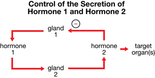
SC 25. If gland 1 is the pituitary gland, the row that identifies hormone 1, gland 2, and hormone 2 is
Row |
Hormone 1 |
Gland 2 |
Hormone 2 |
A. |
ACTH |
adrenal cortex |
adrenaline |
B. |
TSH |
thyroid |
thyroxine |
C. |
ACTH |
adrenal medulla |
cortisol |
D. |
ADH |
kidney |
aldosterone |
Use the following information to answer question SC 26.
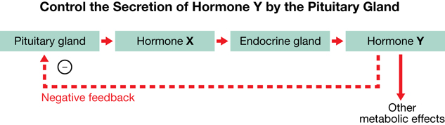
SC 26. Normally, inhibition of the pituitary gland would occur if the secretion of hormone X
- increased, causing a decrease in the secretion of hormone Y
- decreased, causing a decrease in the secretion of hormone Y
- increased, causing an increase in the secretion of hormone Y
- decreased, causing an increase in the secretion of hormone Y
 Self-Check Answers
SC 16. B
SC 17. C
SC 18. C
SC 19. D
SC 20. A
SC 21. B
SC 22. D
SC 23. A
SC 24. D
SC 25. B
SC 26. C
Reflect on the Big Picture
The endocrine system is self-regulating through its boss and through negative feedback mechanisms. You have had the opportunity to reflect on which part of the hypothalamus-pituitary complex you think is in charge. You have discussed your choice with your peers and teacher. But what happens when the boss of the endocrine system is not doing its job, as in Emily’s case? How are the regulatory pathways affected when tropic hormones are not functioning properly or aren’t present in adequate amounts?
In Lesson 3 you will examine Emily’s inability to regulate the stress hormones produced by the adrenal gland because her pituitary tumour causes too much ACTH to be secreted. In Lesson 4 you will examine the effects of her body not being able to regulate the thyroid hormone thyroxine because the tumour causes the undersecretion of TSH.
 Module 2: Lesson 2 Assignment
Submit your completed Module 2: Lesson 2 Assignment to your teacher for assessment.
1.19. Page 8
Module 2—The Endocrine System
 Lesson Summary
In this lesson you explored the following focusing question:
- Who is the boss of the endocrine system, and how is the control of the endocrine system managed?
Self-regulated by the hypothalamus-pituitary complex, the endocrine system utilizes tropic hormones, such as releasing hormones from the hypothalamus and ACTH, TSH, and hGH from the anterior pituitary, to stimulate other endocrine glands, such as the adrenal gland and thyroid gland, to produce their hormones. Negative feedback reduces the secretion of the tropic hormones when the levels of hormones secreted by the target glands reach correct amounts. Disruption in the level of a hormone can cause serious effects on physiological functions, such as urine production, growth, and metabolic rate.
You may choose to review the hormones of this lesson by watching all of “The Hypothalamus and Pituitary Gland: The Master Complex.”
Lesson Glossary
Consult the glossary in the textbook for other definitions that you may need to complete your work.
acromegaly: a condition brought about by excessive secretion of human growth hormone in an adult; hands, feet, and nose widen and become enlarged; jaw protrudes; voice becomes husky; barrel chest may develop; sweat glands enlarge; heart enlarges; high blood pressure may develop; enlarged tissues press on nerves, especially the optic nerves, causing loss of vision, particularly in the outer fields; and pressure on the brain causes severe headaches
There is an increased likelihood of developing diabetes mellitus.
anterior pituitary: an endocrine gland consisting of secretory cells that synthesize and secrete several hormones directly into the bloodstream
diabetes insipidus: a condition caused by lack of ADH, which results in excessive production of very dilute urine
ADH may be produced in insufficient quantities by the hypothalamus or the posterior pituitary may fail to release it into the bloodstream when a tumour develops.
gigantism: a condition where a person produces excess hGH during childhood, resulting in a height of 8 feet or more
hypothalamus: the region of the brain located below the cerebral hemispheres and thalamus and just above the pituitary gland; functions in maintaining homeostasis, and is especially important in co-ordinating the endocrine and nervous systems; secretes hormones of the posterior pituitary as well as releases hormones that regulate the anterior pituitary
neurosecretory cells: specialized nerve cells in the hypothalamus that extend into the posterior pituitary and secrete ADH and oxytocin into the posterior pituitary and, subsequently, into the bloodstream
pituitary dwarfism: a condition where a person does not produce enough hGH during childhood, resulting in short stature
posterior pituitary: an extension of the hypothalamus composed of nervous tissue that secretes hormones produced in the hypothalamus into the bloodstream; consists of a temporary storage site for hormones produced in the hypothalamus
releasing hormones: hormones produced by neurosecretory cells in the hypothalamus that stimulate or inhibit the secretion of hormones by the anterior pituitary; sometimes called releasing factors
vasopressin: also called antidiuretic hormone (ADH)
1.20. Lesson 3
Module 2—The Endocrine System
Lesson 3—the Adrenal Gland
 Get Focused
When you studied the sympathetic nervous system in Module 1, you learned about the fight-or-flight response, which refers to the tendency in stressful situations to run away or stay and work through the situation. Imagine watching a horror movie and anticipating the feeling of being scared out of your skin. If your life were a horror movie, you would constantly be on edge—anxious and just waiting to be scared. Life would be a constant adrenaline rush. How long do you think you could last like that?
Emily’s life is like a never-ending horror movie. Tiny tumours in and on her adrenal glands cause the glands to overproduce epinephrine, norepinephrine, and cortisol. These are hormones that combat both short-term and long-term stress. As a result, Emily’s body, left untreated, prepares itself for the fight-or-flight response all the time.
In this lesson you will explore the following focusing question:
- How do the adrenal glands and their secretions affect the body?

 Module 2: Lesson 3 Assignment
Download a copy of the Module 2: Lesson 3 Assignment to your computer now. You will receive further instructions about how to complete this assignment later in the lesson.
You must decide what to do with the questions that are not marked by the teacher.
Remember that these questions provide you with the practice and feedback that you need to successfully complete this course. You should respond to all the questions and place those answers in your course folder.
The assignment that you will submit to your teacher for assessment involves questions about the adrenal gland and homeostasis. In your response you will use feedback loops, flow charts, tables, and Venn diagrams.
This lesson is devoted entirely to the study of the adrenal gland. The concepts are significant in Biology 30. While you are completing this lesson, there will be many opportunities for you to acquire, understand, and practise the concepts that are presented to you. As you complete these activities, as well as your summary notes, you will file everything in your course folder to reference when you are preparing for exams.
Remember you also have the option of trying additional questions from the textbook for further practice. Consult with your teacher for the answers to these questions. The Key will also provide you with many Diploma Exam-style multiple-choice, numerical-response, and written-response questions that will be an excellent review of the module. Practising your responses to these types of questions is good preparation for the Diploma Exam.
1.21. Page 2
Module 2—The Endocrine System
 Explore
Adrenal Gland and Stress Reactions
 Read
Read

© Vladimir Mucibabic/shutterstock
If you look at “Figure 13.19(B)” on page 451 of the textbook, you will see that the adrenal glands look like small baseball caps perched on top of the kidneys. Further examination shows that each adrenal gland is really two glands with the adrenal cortex (meaning “bark”) wrapped around the adrenal medulla (meaning “middle”). The adrenal cortex consists of layers of secretory cells, whereas the adrenal medulla consists of modified nerve cells. What other gland have you studied that is made of both nervous and endocrine tissue? If you answered, “the pituitary gland,” you are correct. Both parts of the adrenal gland produce and release hormones in response to stress stimuli.
In the Student Stress lab that you completed in Lesson 1, you created a stressful situation for your partner. The adrenal gland would have played a significant role in bringing about changes in your partner’s blood pressure and heart rate. The stress reactions of the human body comprise a wide range of reactions that prepare the body to resist any damaging or potentially damaging changes in the environment. A great variety of stimuli trigger these responses, including injury, infection, and emotional responses, such as fear or anger. The effectiveness of a particular stimulus varies from person to person. You may be freaking out about that exam in the afternoon, but your friend may not be worried about it at all. Or the bass in your car stereo may be driving your parents nuts, but you’re enjoying the beat.
adrenal cortex: the outer portion of the adrenal gland composed of three distinct layers of cells that respond to hormonal signals from the anterior pituitary by producing three groups of hormones, including the glucocorticoids, mineral corticoids, and gonadocorticoids; reacts to stress stimuli and affects salt and water balance
adrenal medulla: the central or middle part of the adrenal gland composed of modified nervous tissue that responds to sympathetic nerve signals by secreting the fight-or-flight hormones, epinephrine and norepinephrine, in reaction to stress
For an introduction to the adrenal glands, read page 451 in your textbook.
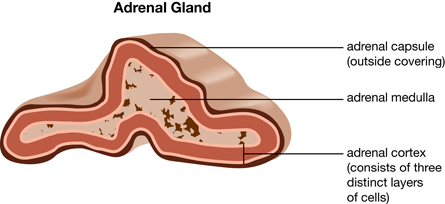
 Watch and Listen
Review and reinforce concepts about the adrenal gland and stress responses by viewing the following two segments of “The Adrenal Glands and the Gonads: Stress and Sexuality: Vital to Life.” You may be required to enter a username and password to access these videos. Contact your teacher for this information.
- “Adrenal Glands”
- “Bio Review: Adrenal Glands and Stress”
1.22. Page 3
Module 2—The Endocrine System
The Adrenal Medulla—Regulating Short-Term Stress
 Read
Read

© John Bell/shutterstock
In Module 1 you studied sympathetic nerve activity, which is collectively called the fight-or-flight responses. These responses prepare the body for immediate action. To refresh your memory and summarize what these responses are, read “The Autonomic System” on page 397 and “The Adrenal Medulla: Regulating the Short-Term Stress Response” on page 452 and the top of page 453 in your textbook.
Nerve impulses from the sympathetic nervous system stimulate the adrenal medulla to release epinephrine and norepinephrine. These hormones circulate in the bloodstream and enhance the activities of the sympathetic nervous system. Together, the sympathetic nervous system and the release of hormones from the adrenal medulla provide the resources (oxygen and blood glucose) for energy release by aerobic cellular respiration and allow the body to perform far beyond its capacity. You may have heard stories of how a mother fearlessly fought off a bear that was mauling her child or how a man lifted a car to free an accident victim pinned beneath it. These incredible feats were performed because the sympathetic nervous system stimulated the adrenal medulla to release a burst of adrenaline, which enabled a lot of energy to be released by metabolism.
fight-or-flight response: a short-term stress response that ensures increased amounts of glucose and oxygen are available to muscle cells for cellular respiration, which results in increased available energy for use by muscles
epinephrine: a hormone produced by the adrenal medulla in response to stress; also called adrenaline
The adrenal gland produces about 80% epinephrine and 20% norepinephrine.
norepinephrine: a second hormone produced by the adrenal medulla in response to stress, but in much smaller quantities than epinephrine
About 20% of adrenal medulla secretions are norepinephrine.
Use the information on page 452 of your textbook to make notes about the adrenal medulla, or prepare a flow chart similar to “Figure 13.21” on page 452. You may also choose to make summary notes from the following video segments.
 Watch and Listen
The following segments of “The Adrenal Glands and the Gonads: Stress and Sexuality: Vital to Life” will reinforce your understanding of how the adrenal medulla works to combat the effects of what is sometimes called short-term stress. You may be required to enter a username and password to access these videos. Contact your teacher for this information.
- “Epinephrine Release”
- “Bio Quest: Fight-or-Flight”
- “Bio Discovery: Body Responses to Epinephrine”
- “Symptoms of Stress”
 Self-Check
To review the concepts of the short-term stress response, complete the following questions. After checking your answers, file them in your course folder.
SC 1. Describe a “fight” response and a “flight” response, and include the biological mechanism behind these responses.
SC 2. Identify three groups of stress stimuli (categories of things that cause stress).
SC 3. Explain how the hypothalamus is involved in the release of epinephrine and norepinephrine by the adrenal medulla. You may choose to use a flow chart to illustrate your response.
Self-Check Answers
SC 1. The short-term responses to stress are commonly referred to as the fight-or-flight responses. Like the nerve impulses of the sympathetic nervous system, the hormones of the adrenal medulla prepare the body to fight or flight by increasing the levels of oxygen and glucose and their distribution to the muscles and tissues. To increase the level of oxygen in the blood, the breathing rate increases and the bronchioles dilate go get more oxygen into the lungs for diffusion. To increase the level of glucose in the blood, the liver and muscles are stimulated to convert stored glycogen into glucose. Heart rate, cardiac output, and blood pressure all increase so that distribution of the blood to the tissues is greater and faster. Peripheral blood vessels (in the skin) and those to the digestive system constrict and redirect blood to the muscles and tissues that need more blood. Cellular respiration is increased and releases more energy to deal with the situation or to quickly run away. In addition, sweating increases to deal with the extra heat produced from increased metabolism (cellular respiration) and the pupils dilate so more light enters the eyeball and increases visual acuity so that the danger can be seen more clearly.
SC 2. Three groups of stress stimuli may be infection; injury to the body; or emotional stimuli, such as fear, anger, or extreme sadness.
SC 3. The adrenal medulla is composed of modified neurons. Neurons from the sympathetic nervous system of the autonomic nervous system, whose centres are located in the hypothalamus, directly stimulate the cells of the adrenal medulla to produce epinephrine and norepinephrine. This can also be explained with a flow chart that may resemble the following sample response.

1.23. Page 4
Module 2—The Endocrine System
The Adrenal Cortex—Regulating Long-Term Stress
 Read
Read

© Fouquin/shutterstock
Cortisol, which is produced by the adrenal cortex, is thought to be the most important glucocorticoid in humans. It is primarily concerned with glucose metabolism during periods of long-term stress, such as engaging in athletic competition and writing Diploma Exams. It also plays a role in increasing blood pressure when it works in concert with the sympathetic nervous system and the release of epinephrine and norepinephrine by the adrenal medulla. After a time, the stress responses fade, and normal conditions are restored in part by the parasympathetic nervous system. In the Student Stress lab your partner slowly returned to normal after you had stressed him or her out. If the stress-producing stimuli persist, the danger to the body can include an excessively high metabolic rate, high blood pressure, and an elevated heart rate for a prolonged period of time—or long-term stress responses. This is particularly damaging for the circulatory system and is a contributing factor in increasing the risk of heart attacks and atherosclerosis, which you studied in Biology 20.
cortisol: also called hydrocortisone or cortisone; involved in enhancing short-term stress responses and in stimulating conversion of protein and fat into glucose
Cortisol is secreted in highest amounts in humans and is produced by the adrenal cortex in response to stimulation by ACTH.
glucocorticoids: a group of hormones produced by the adrenal cortex, of which cortisol is of prime importance; influence glucose metabolism from protein and fat and immune function by suppressing inflammation
long-term stress responses: responses, mainly stimulated by cortisol, which are of long duration
Cortisol also acts to suppress inflammation, and is sometimes used in the treatment of arthritis and allergies. Side effects, such as reducing immune system effectiveness, limit its usefulness.
You can further your understanding of the role that cortisol plays in returning the body to homeostasis by reading pages 453 and 454 of your textbook or by watching the following video segments of “The Adrenal Glands and the Gonads: Stress and Sexuality: Vital to Life.” Whichever you choose to do, make notes to support your understanding and file them in your course folder for later review.
If you choose to watch the videos, you may be required to enter a username and password to access the videos. Contact your teacher for this information.
- “Adrenal Cortex”
- “Bio Fact: Glucocorticoid Hormones”
Regulating Cortisol Release
Cortisol is secreted in response to ACTH when the anterior pituitary is stimulated by releasing hormone from the hypothalamus. Secretion is decreased by negative feedback exerted on the anterior pituitary and the hypothalamus, as outlined in the general regulatory pathway of tropic hormones in “Figure 13.10” on page 441 of your textbook and in the following segment of “The Adrenal Glands and the Gonads: Stress and Sexuality: Vital to Life.”
- “Adrenocorticotropic Hormone”
This is one of the more simple feedback loops that you will study in this module. The information that you have learned about cortisol could be easily summarized in a well-labelled feedback loop diagram for your course folder.
 Self-Check
Use the following information to answer the next four questions, remembering that your responses must include well-explained steps that follow a sequential order of the events. After checking your answers, file them in your course folder. Consult with your teacher if you do not understand any questions or answers or if you need feedback about the detail of your responses.
| Due to tiny tumours growing in Emily’s anterior pituitary, she overproduces ACTH, which, in turn, stimulates oversecretion of cortisol. The cortisol maintains a high metabolic rate by burning protein and fat; constricts her blood vessels, causing an increase in her blood pressure; and causes her heart to race constantly. |
SC 4. Draw a feedback loop that would illustrate this disruption in Emily’s production of cortisol.
SC 5. Before treatment, Emily’s blood glucose was abnormally high. Explain how this situation could have been the result of overproduction of cortisol.
SC 6. Emily’s heart rate and blood pressure were elevated around the clock. Explain why this is a potentially life-threatening situation.
SC 7. Emily was very prone to infections, such as colds, laryngitis, and bronchitis. Explain why this was probably the result of elevated cortisol levels.
Self-Check Answers
SC 4. The feedback loop should illustrate the positive feedback in the tropic regulatory pathway for cortisol and aldosterone. It should resemble the one shown below, but could be drawn horizontally rather than vertically. Boxes could be placed around the words hypothalamus, anterior pituitary, and adrenal cortex.
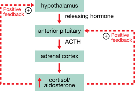
SC 5. Cortisol stimulates the decomposition of proteins, amino acids, and fats, as well as the conversion of the products of their breakdown into glucose. Too much cortisol would accelerate this process and explain why Emily’s blood had an elevated glucose level.
SC 6. An increased heart rate and high blood pressure lead to increased risk of heart attack, stroke, aneurysms (bursting of blood vessels), and kidney damage, which are all life-threatening situations.
SC 7. Inflammation is the immune system’s response to injury and invasion by micro-organisms. It directs the body’s defences to block off the area, kill the invaders, dispose of dead and damaged tissue, and begin the process of repair. During inflammation, the blood supply to the area is increased and the area becomes red and warm. The walls of the blood vessels become leaky and the area swells. White blood cells flock to the area to kill the bacteria or viruses that are present, and they release chemicals that continue the inflammation. Cortisol decreases the inflammatory response. If the inflammatory response is decreased by elevated levels of cortisol, the invading micro-organisms are not killed and the person becomes sick with a cold, laryngitis, bronchitis, pneumonia, and so on. Also, the person may become ill more often if the inflammatory response is dampened.
1.24. Page 5
Module 2—The Endocrine System

© Peter Miller/iStockphoto
Aldosterone
 Read
Read
A second group of hormones produced by the adrenal cortex is called the mineralocorticoids, which include aldosterone. In Biology 20 you learned that aldosterone targets the distal and collecting tubules of the kidneys, resulting in an increase in the absorption of sodium into the blood. To review the role of aldosterone and how it contributes to maintaining homeostasis, read “Aldosterone” on page 454 in your textbook. Take point-form notes for your course folder as you read.
mineralocorticoid: a hormone produced by the adrenal cortex that regulates salt and water homeostasis by influencing the reabsorption of sodium ions
An example is aldosterone.
aldosterone: a hormone produced by the adrenal cortex in response to an increase in certain enzymes and high levels of ACTH; acts to increase sodium ion reabsorption
 Self-Check
To apply your understanding of how aldosterone contributes to homeostasis involving sodium ions and water, complete the questions SC 8, SC 9, and SC 10 using full sentences.
SC 8. What is the difference in the way that aldosterone and ADH affect water balance?
SC 9. Why are sodium ions important in the body?
SC 10. How does aldosterone help the body cope with an ongoing stressful situation? How is this response different from the fight-or-flight response?
Complete the following questions in preparation for the Lesson 3 Assignment. These questions consist of both multiple-choice and written-response questions. When you have finished, check your answers. If you do not understand any of the concepts, be sure to discuss them with your teacher.
SC 11. Increasing the levels of aldosterone in the blood would result in
- increased blood volume
- decreased blood pressure
- decreased heart rate
- increased urine production
SC 12. During an emergency situation, the adrenal gland is stimulated to release a hormone that directly causes an increase in
- sodium ion concentration
- blood glucose levels
- conversion of glucose to glycogen
- sympathetic nervous stimulation
SC 13. Returning involuntary body functions to normal after a period of stress is partly the function of the
- sympathetic nervous system
- parasympathetic nervous system
- tropic hormones such as ACTH
- releasing hormones from the hypothalamus
SC 14. A high blood cortisol level controls the secretion of
- adrenocorticotropic hormone and releasing hormone from the hypothalamus
- adrenocorticotropic hormone and releasing hormone from the anterior pituitary
- adrenocorticotropic hormone from the hypothalamus and releasing hormone from the anterior pituitary
- adrenocorticotropic hormone from the anterior pituitary and releasing hormone from the hypothalamus
SC 15. Which of the following is normally not part of the fight-or-flight response to short-term stress situations?
- increased blood glucose levels
- increased heart rate
- increased blood pressure
- increased blood volume
Use the following information to answer questions SC 16 and SC 17.
| Polygraphs (lie detectors) monitor certain changes in some activities of the nervous system. In theory, an emotionally stressful situation, like telling lies, will increase a person's perspiration, breathing rate, and heart rate and cause slight dilation of the pupils. However, polygraphs cannot exclusively differentiate between telling lies and other stressful situations. |
SC 16. The physiological changes that are associated with telling lies are responses produced by
- impulses from sympathetic nerves of the autonomic nervous system and hormones from the adrenal medulla
- impulses from sympathetic nerves of the autonomic nervous system and hormones from the adrenal cortex
- impulses from parasympathetic nerves of the autonomic nervous system and hormones from the adrenal medulla
- impulses from the parasympathetic nerves of the autonomic nervous system and hormones from both the adrenal medulla and the cortex
SC 17. Emotionally stressful situations may affect more than one system of the body. Another possible response produced by telling lies would be
- decreased secretion of ACTH
- increased secretion of aldosterone
- decreased secretion of cortisol
- increased secretion of epinephrine
Use the following information to answer question SC 18 and SC 19.
| Stressful situations trigger the release of hormones such as cortisol. Recent studies have found that some forms of depression cause a similar hormonal response that lasts much longer than the normal stress response. This unregulated release of stress hormones may result in reduced appetite, an unresponsive immune system, inadequate tissue repair, and insomnia. |
SC 18. Another stress hormone whose functions mimic those of the sympathetic nervous system is
- acetylcholine
- norepinephrine
- aldosterone
- insulin
Use the following additional information to answer the next question.
SC 19. A logical interpretation of the graph is that the
- secretion of cortisol is inhibited by increased ACTH
- secretion of cortisol is doubled if the secretion of ACTH is doubled
- adrenal glands respond more quickly to small amounts of ACTH than to large amounts of ACTH
- adrenal glands respond to large amounts of ACTH by having a maximum cortisol secretion rate
SC 20. The secretion of Hormone A causes an increase in Activity X in the body. If this mechanism works by positive feedback, which of the following statements is true?
- An increase in X produces an increase in A.
- An increase in A produces a decrease in X.
- An increase in X produces a decrease in A.
- A decrease in A produces an increase in X.
SC 21. Suppose you are lost in concentration while studying biology. Suddenly, the phone rings and startles you. Outline the physiological changes that occur in your body due to release of the stress hormones cortisol and epinephrine. What triggers the release of each of these hormones?
SC 22. The appropriate level of hormones circulating in the body is critical to proper homeostasis. If there is too much cortisol and aldosterone, a person develops Cushing’s disease. If there is too little of these hormones, a person develops Addison’s disease. Complete a table like the one below contrasting the two disorders
Contrasting Oversecretion and Undersecretion of Adrenal Cortex Hormones
Features |
Cushing’s Disease |
Addison’s Disease |
Hormones Involved |
|
|
Level of Hormones Secreted |
|
|
Cause of Inappropriate Level of Secretion |
|
|
Symptoms |
|
|
Treatment |
Self-Check Answers
SC 8. Aldosterone first increases reabsorption of sodium ions into the blood from the kidneys, causing an increase in solute concentration of the blood, which results in water reabsorption. ADH directly increases the permeability of cells in the nephrons of the kidneys to reabsorb more water.
SC 9. Sodium ions are very important in nerve impulse transmission (studied in Module 1), muscle contraction, and ion (electrolyte) balance in the body.
SC 10. Aldosterone targets the kidneys, where it promotes the renal absorption of sodium ions into the bloodstream. The resulting increase in the solute concentration of the blood draws more water from the nephrons in the kidneys, resulting in higher blood pressure. Higher blood pressure would be advantageous as a stress response because more oxygen and glucose from the blood would be made available to the tissues in less time. However, this response would take place over a much longer period of time than the fight-or-flight response and may prove to be harmful to the circulatory system if sustained for such long periods of time, resulting in increased risk of heart attack and atherosclerosis.
SC 11. A
SC 12. D
SC 13. B
SC 14. D
SC 15. D
SC 16. A
SC 17. D
SC 18. B
SC 19. D
SC 20. A
SC 21. When you are startled, the adrenal medulla is stimulated by neurons from the sympathetic nervous system to release the hormones epinephrine and norepinephrine. These hormones trigger an increase in breathing rate, heart rate, blood pressure, blood flow to the heart and muscles, and the conversion of glycogen to glucose in the liver. In other words, these hormones trigger all of the fight-or-flight responses. In addition, when the brain detects danger, it signals the hypothalamus to secrete a releasing hormone. The releasing hormone stimulates the anterior pituitary gland to secrete ACTH. ACTH stimulates the adrenal cortex to release cortisol. Cortisol intensifies the responses of epinephrine and norepinephrine, but is longer lasting. Cortisol also increases blood glucose by promoting the breakdown of proteins (amino acids) and fats to glucose (not by conversion of glycogen to glucose like epinephrine and norepinephrine).
SC 22. The completed table should have the following differences.
Features |
Cushing’s Disease |
Addison’s Disease |
Hormones Involved |
cortisol, aldosterone |
cortisol, aldosterone |
Level of Hormones Secreted |
too much |
too little |
Cause of Inappropriate Level of |
tumours in anterior pituitary, causing oversecretion of ACTH
OR tumours in adrenal cortex, causing oversecretion of cortisol and/or aldosterone |
destruction of cells in adrenal cortex (auto-immune disease) |
Symptoms |
excess accumulation of fat at back of neck (buffalo hump) or in puffiness of the face, causing a round or “moon” face, increased blood pressure, and decreased resistance to infections |
sodium ions not balanced, loss of water in urine, low blood glucose, decreased blood pressure |
Treatment |
surgery to remove tumours or radiation to shrink tumours, drugs to dampen effects of ACTH |
cortisol in the form of prednisone or hydrocortisone by mouth in tablet form |
Based on your success with the Self-Check, you may wish to practise some of the questions on page 455 of your textbook. Discuss your responses with your teacher.
 Module 2: Lesson 3 Assignment
Retrieve the copy of the Module 2: Lesson 3 Assignment that you saved to your computer earlier in this lesson. Complete all questions. Save your completed assignment in your course folder. You will receive instructions about when to submit your assignment to your teacher later in this lesson.
1.25. Page 6
Module 2—The Endocrine System
 Reflect and Connect
Stress, in moderate doses, is a good thing! Based on the concepts that you have reviewed in this lesson, reflect on how stress is good. What kinds of activities are carried out better under some stress? Do you think athletes would do as well if they did not experience the stress of competition that gives them that extra burst of epinephrine? If your heart did not beat a little faster and your breathing rate was not increased at all, do you think you would do as well on an exam? Why do you think “extreme sports” are so popular? Is being too laid-back a good thing in some situations? How is too much stress bad for you? Why does a constant accelerated heart rate and constant higher-than-normal blood pressure lead to heart attacks? Reflect on these questions, and use the concepts that you have learned in this lesson to formulate possible responses. Share your responses in the class discussion area. Did any of the other students surprise you with their response?
Reflect on the Big Picture
Due to FMEN 1, Emily has developed tumours in both her anterior pituitary and in her adrenal cortex. How has this affected the secretion of hormones in her body? What has happened to her stress responses? If the tumours had not been diagnosed and treated, what potentially life-threatening risks might she have faced?
 Going Beyond
Hans Selye, a scientist at the University of Montreal, first described the general adaptation syndrome (GAS). GAS has three main stages: alarm, resistance, and exhaustion. However, Selye did not regard all stress as bad, and even said that some stress was the “spice of life.” Researchers have called this good stress “eustress.” Using library sources as well as the Internet, research GAS.
 Module 2: Lesson 3 Assignment
Submit your completed Module 2: Lesson 3 Assignment to your teacher for assessment.
1.26. Page 7
Module 2—The Endocrine System
 Lesson Summary
The following focusing question was explored in this lessson:
- How do the adrenal glands and their secretions affect the body?
Located atop the kidneys, the adrenal gland is really two glands. Similar to the two lobes of the pituitary gland, the adrenal cortex is composed of several layers of secretory cells, while the adrenal medulla is made up of modified nerve cells. Hormones from both parts of the adrenal gland deal with stress. Epinephrine and norepinephrine from the adrenal medulla intensify the fight-or-flight responses of the sympathetic nervous system. The purpose of these responses is to get more oxygen and glucose to the tissues. For example, breathing rate increases and the bronchioles dilate so more oxygen can be taken to the lungs. Glucose is synthesized from glycogen. Heart rate, cardiac output, and blood pressure are increased so that the increased levels of oxygen and glucose can be delivered to the muscles. Increased cellular metabolism releasing energy in the muscles then makes it possible to “fight or flight.” Cortisol, from the adrenal cortex, intensifies the fight-or-flight responses. It increases blood glucose by converting amino acids, proteins, and fats into glucose. Cortisol also decreases the inflammatory response, so it can be useful in treating allergic reactions and joint inflammations. High levels of cortisol are regulated by negative feedback on the hypothalamus and anterior pituitary and their secretions of releasing hormone and ACTH, respectively. Aldosterone, another hormone from the adrenal cortex, is concerned with regulation of sodium ions and subsequent homeostasis of water in the tissues.
 Watch and Listen
“The Adrenal Glands and the Gonads: Stress and Sexuality: Vital to Life” provides an excellent review of the concepts of this unit and includes many technologies and societal issues. You may choose to view the entire video at this point.
Lesson Glossary
Consult the glossary in the textbook for other definitions that you may need to complete your work.
adrenal cortex: the outer portion of the adrenal gland composed of three distinct layers of cells that respond to hormonal signals from the anterior pituitary by producing three groups of hormones, including the glucocorticoids, mineral corticoids, and gonadocorticoids; reacts to stress stimuli and affects salt and water balance
adrenal medulla: the central or middle part of the adrenal gland composed of modified nervous tissue that responds to sympathetic nerve signals by secreting the fight-or-flight hormones, epinephrine and norepinephrine, in reaction to stress
aldosterone: a hormone produced by the adrenal cortex in response to an increase in certain enzymes and high levels of ACTH; acts to increase sodium ion reabsorption
cortisol: also called hydrocortisone or cortisone; involved in enhancing short-term stress responses and in stimulating conversion of protein and fat into glucose
Cortisol is produced by the adrenal cortex in response to stimulation by ACTH.
epinephrine: a hormone produced by the adrenal medulla in response to stress; also called adrenaline
The adrenal gland produces about 80% epinephrine and 20% norepinephrine.
fight-or-flight response: a short-term stress response that ensures increased amounts of glucose and oxygen are available to muscle cells for cellular respiration, which results in increased available energy for use by muscles
glucocorticoids: a group of hormones produced by the adrenal cortex, of which cortisol is of prime importance; influence glucose metabolism from protein and fat and immune function by suppressing inflammation
long-term stress responses: responses, mainly stimulated by cortisol, which are of long duration
mineralocorticoid: a hormone produced by the adrenal cortex that regulates salt and water homeostasis by influencing the reabsorption of sodium ions
norepinephrine: a second hormone produced by the adrenal medulla in response to stress, but in much smaller quantities than epinephrine
About 20% of adrenal medulla secretions are norepinephrine.
1.27. Lesson 4
Module 2—The Endocrine System
Lesson 4—the Thyroid and Parathyroid Glands
 Get Focused

© mammamaart/iStockphoto

© Ed Hidden/istockphoto
Hyperthyroidism and hypothyroidism are two opposite types of thyroid disorders. If you have hyperthyroidism, you might always feel too hot, experience weight loss, and find it difficult to concentrate. You may also be jittery and hyperactive. If you have hypothyroidism, you are likely to feel cold, you may be overweight, you may feel lethargic, and you may have trouble staying awake. Emily developed hypothyroidism with her condition. She also developed tiny growths in the parathyroid glands. An overproduction of a parathyroid hormone caused her to lose calcium from her bones, which became fragile and brittle and caused her to develop kidney stones.
In this lesson you will explore the following focusing question:
- How do the thyroid and parathyroid glands contribute to homeostasis?
 Module 2: Lesson 4 Assignment
Download a copy of the Module 2: Lesson 4 Assignment to your computer now. You will receive further instructions about how to complete this assignment later in the lesson.
You must decide what to do with the questions that are not marked by the teacher.
Remember that these questions provide you with the practice and feedback that you need to successfully complete this course. You should respond to all the questions and place those answers in your course folder.
While you are completing this lesson, there will be many opportunities for you to acquire, understand, and practise the concepts that are presented to you. As you complete these activities, as well as your summary notes, you will file everything in your course folder to reference when you are preparing for exams.
Remember you also have the option of trying additional questions from the textbook for further practice. Consult with your teacher for the answers to these questions. The Key will also provide you with many Diploma Exam-style multiple-choice, numerical-response, and written-response questions that will be an excellent review of the module. Practising your responses to these types of questions is good preparation for the Diploma Exam.
1.28. Page 2
Module 2—The Endocrine System
 Explore
Anatomy of the Thyroid Gland
 Read
Read
© Sebastian Kaulitzki/shutterstock
The structure of the thyroid gland is different from other endocrine glands. Thyroid tissue contains many spherical follicles consisting of a single layer of secretory cells surrounding a storage space. The secretory cells produce a precursor of thyroxine, which is stored in the storage space. Stimulation by thyroid stimulating hormone, which is produced by the anterior pituitary, causes the follicles to change the precursor and release it as thyroxine into the many blood capillaries between the follicles. Look at “Figure 13.16(C),” a micrograph of thyroid tissue, on page 448 of your textbook. Notice that there are some large cells (C cells or parafollicular cells) between the follicles. These cells produce and secrete calcitonin, another important thyroid hormone that will be addressed later in this lesson.
follicle: as it relates to the thyroid gland, a microscopic structure consisting of a circle of cells called follicle cells surrounding a central cavity
The thyroid gland consists of many follicles.
precursor: an inactive form of a molecule that can easily be changed into the active form
The precursor thyroglobin can easily be changed into the active thyroxine by the addition of iodine atoms.
thyroxine: a hormone produced by the follicle cells of the thyroid gland; mainly controls the rate at which the body metabolizes carbohydrates, fats, and proteins for energy and stimulates the proper development of the nervous system
C cells (parafollicular cells): cells located between the follicles of the thyroid gland that are responsible for synthesizing and secreting calcitonin
calcitonin: a hormone produced by the C cells (parafollicular cells) in the thyroid gland that decreases blood calcium levels by increasing uptake of calcium into the skeleton and inhibiting decomposition of bone when the blood levels of calcium are too high
Read “The Thyroid Gland: A Metabolic Thermostat” on pages 446 and 447 in your textbook. Study “Figure 13.16” on page 448, and then complete the following Self-Check activity.
 Self-Check
SC 1. On the diagram of thyroid tissues, label the follicle, storage space, C cells, and blood capillaries (vessels). Annotate the diagram by indicating the function of each structure, in brackets, after its label. Once you have finished, check your diagram and annotations and store it in your course folder.
Self-Check Answers
SC 1.
1.29. Page 3
Module 2—The Endocrine System
The Thyroid Gland and Metabolism
 Read
Read
metabolism: all of the chemical reactions that occur in living organisms to support and sustain life processes
One type involves the buildup or synthesis of complex compounds from simple ones. A second type consists of the breakdown or decomposition of complex compounds into simple ones. Cellular respiration is one of the major metabolic processes.
cellular respiration: a process occurring outside and inside the mitochondria of cells of all living organisms in which energy-rich compounds, such as carbohydrates (glucose), proteins, and fats, are burned with oxygen to release energy in the form of ATP and waste products, such as water and carbon dioxide
Do you know someone who seems to be able to consume great amounts of food and still stay thin? Do you know other people who seem to put on weight just by smelling or looking at food? Thin people often say they have a high metabolism. Metabolic rate is regulated by the hypothalamus-pituitary complex, which produces tropic hormones that stimulate the thyroid gland to release increased amounts of thyroxine. Thyroxine increases the number of mitochondria in cells and generally increases cellular respiration, a major set of decomposition chemical reactions that make up metabolism. You may wish to review these concepts from Biology 20 by looking through “Section 5.3: Cellular Respiration Releases Energy from Organic Compounds,” which begins on page 182 of your textbook.
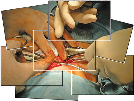
© Carolina K. Smith, M.D./shutterstock
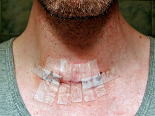
![]() Watch and Listen
Watch and Listen
To gain an understanding of the composition of thyroxine, how it is regulated, what functions it performs, and what happens when it is oversecreted or undersecreted, watch “The Thyroid Gland: Waging the Battle for Metabolism.” Some significant sections of this video are listed below. It is interesting to view the video to see the physical consequences of cretinism (hyposecretion) and Grave’s disease (hypersecretion).
- “Thyroid Gland Overview”
- “Bio Fact: Thyroxine”
- “Bio Bit: Cretinism”
- “Bio Discovery: Neonatal Hypothyroidism (Cretinism)”
- “Hypothyroidism”
- “Thyroxine Production”
- “Negative Feedback Loop: Thyroxine”
- “Goiter”
- “Bio Bit: Iodine Storage”
- “Hyperthyroidism”
- “Bio STS: Grave’s Disease”
 Self-Check
To apply your underdstanding, answer the following Self-Check questions. The completed answers to these questions can become your notes for this lesson, so it is important to answer in complete sentences. To help you answer the questions, you may use library resources, the Internet, pages 446 through 448 in your textbook, or any of the video segments that you watched. Check your work carefully and file your work in your course folder. Consult with your teacher if you need any clarification.
Use the following information to answer the questions that follow.
| Usually people who have FMEN1 do not develop tumours in the thyroid gland, and Emily did not have a thyroid tumour. However, in Lessons 2 and 3, you learned that Emily had tiny tumours in her anterior pituitary, which affected the tropic regulatory pathway for ACTH by causing the oversecretion of this hormone. Those tumours have also affected the secretion of thyroid stimulating hormone (TSH) by causing an undersecretion of this regulatory hormone. |
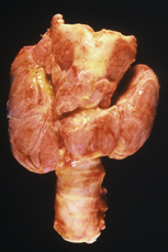
SC 2. Draw a feedback loop that would illustrate the proper negative feedback regulation on the production of thyroxine.
SC 3. Explain how Emily’s feedback loop would be different from the one you drew in question SC 2.
SC 4. Identify the element that is necessary for the synthesis of thyroxine. Although you may live in a zone where this element is in short supply in the soil and water, you are able to produce a fully functioning thyroxine. Explain why.
SC 5. Outline the functions of thyroxine in both children and adults.
SC 6. Emily’s feedback mechanism was not working, and she could not produce adequate amounts of thyroxine. Identify Emily’s resulting thyroid disorder.
SC 7. Describe the symptoms that Emily would exhibit as a result of a lack of thyroxine. Outline a possible treatment for Emily.
SC 8. Define goitre. Left untreated, do you think Emily could have developed a goiter? Explain why or why not.
SC 9. Outline the results of undersecretion of thyroxine in newborn babies. Identify this disorder.
SC 10. Due to the anterior pituitary tumour, Emily could have just as easily oversecreted thyroxine as she had undersecreted it. Describe the symptoms of oversecretion of thyroxine. Identify the disorder associated with the oversecretion of thyroxine and outline a possible treatment.
SC 11. Is hyperthyroidism different from Grave’s disease? Explain.
Self-Check Answers
SC 2. The negative feedback loop regulating thyroxine in the blood should resemble the one shown below. High levels of thyroxine inhibit the hypothalamus from secreting the releasing hormone (which is sometimes abbreviated as TRH—thyroid releasing hormone) and, thus, do not allow stimulation of the anterior pituitary. It also inhibits the anterior pituitary directly by limiting TSH production. You may have placed boxes around the words hypothalamus, anterior pituitary, and thyroid gland, but it is not necessary. The loop may be drawn horizontally rather than vertically.
Negative Feedback Regulation of Thyroxine

SC 3. Because of the tumour in Emily’s anterior pituitary, she does not secrete the proper amount of TSH. Thus, her thyroid gland does not produce enough thyroxine, and the low levels of thyroxine constantly stimulate both the hypothalamus and the anterior pituitary. Since the tumours prevent the secretion of TSH, the stimulation is relentless.
SC 4. Iodine is the element necessary for the production of thyroxine. To ensure adequate iodine in the human diet, it is put into table salt as potassium iodide (K2I). Also, the udders of dairy cows are disinfected with an iodine solution before milking and a small amount of iodine is present in dairy products.
SC 5. Thyroxine influences almost every tissue in the body in order to increase the rate at which the cells metabolize fats, proteins, and carbohydrates for energy purposes. It especially stimulates the heart, skeletal muscles, liver, and kidneys to increase their rate of cellular respiration. Therefore, in adults, thyroxine affects heart rate, respiratory rate, the rate at which calories are burned, skin maintenance, heat production, and it promotes normal adult nervous system function. In children and infants, thyroxine is particularly important in stimulating growth and development of the skeleton and development of the nervous system.
SC 6. Lack of thyroxine in the body is called hypothyroidism. A very severe case in adults may be referred to as myxedema.
SC 7. Emily may show any of the following symptoms of hypothyroidism:
- general slowing of body functions
- fatigue
- slow heart rate, decreased blood pressure, and weak pulse
- puffy skin
- hair loss
- weight gain even though she eats little
- slow speech
- inability to tolerate cold
- slow mental function
Treatment usually involves taking thyroxine in pill form.
SC 8. Goitre is an enlargement of the thyroid gland. The enlargement is brought about by the accumulation of the precursor for thyroxine stored in the central cavity. Hypothyroidism (myxedema) usually results in goitre. The anterior pituitary produces TSH, which relentlessly stimulates the thyroid gland to produce thyroxine, but the only result is the accumulation of more and more precursor in the central cavity of the follicles.
Emily would probably not develop a goitre because her diet contained adequate amounts of iodine (iodized salt). Further, she is lacking stimulation of the thyroid gland because she undersecretes TSH and, therefore, her thyroid gland should remain at a normal size.
SC 9. Undersecretion of thyroxine in a baby, called cretinism, results in stunted growth of the skeleton, which causes the child to be short in stature, as well as in poor development of the nervous system, which may result in poor mental development.
SC 10. Oversecretion of thyroxine is called hyperthyroidism, and it generally causes a speeding up of body functions. Most people will have an enlarged thyroid gland (exothalmic goiter). Most of the other symptoms reflect the speeding up of body functions, including
- increased heart rate, increased blood pressure, and strong pulse
- excessive sweating
- intolerance to heat
- nervousness and anxiety
- difficulty sleeping
- weight loss in spite of increased appetite
- retention of fluid in eyes causing bulging eyes
Treatment involves the surgical removal of certain active parts of the thyroid gland or the administration of radioactive iodine, which selectively destroys the most active follicle cells. This is possible because of the fact that iodine is automatically taken up by the thyroid gland and does not spread to other body organs.
SC 11. Grave’s disease is one type of hyperthyroidism. It is an autoimmune disorder where the thyroid gland is stimulated to produce excess thyroxine by proteins that mimic TSH. An enlarged thyroid gland and protruding eyeballs are both common symptoms of Grave’s disease.
1.30. Page 4
Module 2—The Endocrine System
Antagonistic Hormones Regulate Calcium Levels
 Read
Read
There are two antagonistic hormones (hormones that work in opposition to one another to establish overall balance or homeostasis) that regulate calcium levels in the blood. One of these hormones, calcitonin, originates from the thyroid gland. The other, parathyroid hormone, originates from the parathyroid glands.
 Self-Check
The following questions will help you to gain an understanding of where these hormones are secreted, how they maintain calcium homeostasis, how their secretion is regulated, and what happens when calcium balance is disrupted. The completed answers to these questions will become your notes for this part of Lesson 4. Therefore, complete your work carefully, and consult with your teacher if you have any questions. You may use library sources, the Internet, page 449 in your textbook, or any other resources to which you have access that will help you complete the questions. Check your answers and file your work in your course folder.
SC 12. Describe at least three functions of calcium in the body.
SC 13. Calcium homeostasis is mainly regulated by two hormones. Identify the two hormones and the glands that produce each of them.
SC 14. The two hormones that you identified in question SC 13 are described as being antagonistic hormones. Define what is meant by this.
SC 15. Describe the mechanism of calcitonin function.
SC 16. Outline the regulation of blood calcium levels by calcitonin.
Use the following additional information to answer questions SC 17, SC 18, SC 19, and SC 21.
| FMEN 1 patients often develop minute tumors in the parathyroid glands. This happened to Emily. Consequently, her parathyroid glands oversecrete parathyroid hormone. |
SC 17. Describe the overall effect of parathyroid hormone. Outline three specific actions on the body caused by this hormone to achieve its overall effect.
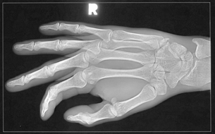
© Troy Kellogg/shutterstock
SC 18. Emily fell on a very icy sidewalk and broke some bones in her wrist. The bones were reset, but months later they still had not healed. During this time Emily was admitted to the emergency room of the hospital with severe abdominal pain, which was diagnosed as a case of kidney stones. Construct a hypothesis that may explain the underlying cause of both health problems. (Remember, a hypothesis should be in the form of an “If . . . then . . . because . . .” statement.)
SC 19. Draw a negative feedback loop that illustrates the regulation of blood calcium levels by parathyroid hormone.
SC 20. How would Emily’s feedback loop be different?
Following are ten multiple-choice questions and two numerical-response questions. Besides being good practice for the type of questions you will encounter on the Diploma Exam, they are good preparation for completing the lesson assessment. Check your answers and, if you do not understand any of the concepts, be sure to discuss them with your teacher.
Multiple-Choice Questions
Use the following diagram to answer questions SC 21 and SC 22.
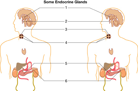
SC 21. Which endocrine gland shown in the illustration would be directly responsible for the development of bone thinning if it was overproducing its hormones?
- 2
- 3
- 4
- 6
SC 22. Cretinism can develop if low secretions of hormone are produced by the gland labelled
- 2
- 3
- 4
- both 2 and 4
SC 23. Hypothyroidism developed in childhood would be characterized by
- impaired physical and mental development
- thirst and consumption of large volumes of water
- increased blood pressure and increased heart rate
- suppression of the immune system and the development of goitre
SC 24. The area of the brain that normally initiates the secretion of TSH is the
- medulla oblongata
- cerebrum
- pons
- hypothalamus
SC 25. A condition that results in an enlargement of the thyroid gland may be caused by a diet that is deficient in
- salt
- iron
- iodine
- sodium
Use the following information to answer questions SC 26, SC 27, and SC 28.
| Vegetables such as cabbage, rutabaga, and turnips contain goitrin, a substance that inhibits iodine uptake by the body. |
SC 26. A person with a diet that is high in vegetables containing goitrin may gain weight fairly rapidly. A possible explanation for this weight gain would be
- increased protein metabolism
- decreased blood sugar levels
- increased glycogen release
- decreased metabolic rate
SC 27. The function of which gland would be most affected by goitrin?
- anterior pituitary
- adrenal cortex
- thyroid gland
- parathyroid gland
SC 28. An increase in goitrin consumption would likely cause a person to experience increased
- fatigue
- heart rate
- breathing rate
- blood pressure
Use the following information to answer questions SC 29 and SC 30.
On April 26, 1986, a major accident occurred at a nuclear generating station in Chernobyl. The nuclear explosion dispersed several tons of radioactive iodine, cesium, uranium, and other elements five kilometers into the air.
Radioactivity is extremely damaging to living cells. From Biosphere 2000: Protecting Our Global Environment |
SC 29. As soon as people in Europe realized there had been a nuclear accident, they rushed to buy iodine tablets. What reason would people have for consuming large quantities of iodine?
- Iodine from the tablets, instead of the radioactive iodine, would accumulate in the thyroid.
- Iodine, by negative feedback, blocks the formation of TSH, therefore protecting the thyroid from radioactivity.
- Iodine inhibits cell division, thereby reducing the amount of cellular damage occurring during exposure to radiation.
- Iodine from the tablets could accumulate in the hypothalamus and block radioactivity from killing or altering body cells.
SC 30. In addition to symptoms of radiation poisoning, which symptoms would be expected among people who did not take iodine tablets?
- pancreatic dysfunction and insufficient insulin production
- metabolic dysfunction, resulting in fatigue and weight gain
- increased ACTH secretion and puffiness of face, chest, and abdomen
- pituitary dysfunction resulting in increased hGH secretion and development of gigantism
Numerical-Response Questions
Use the following information to answer question SC 31.
Some Glands and Hormones of the Human Body
Glands
Hormones
|
SC 31. Select a set of numbers for glands and hormones that correctly completes the statement below.
The ______ secretes ______, which stimulates the ______ to produce ______,
(gland) (hormone) (gland) (hormone)
which regulates metabolism (cellular respiration).
Use the following information to answer question SC 32.
Endocrine Glands
|
SC 32. Using the numbers that represent the endocrine glands shown above, match the hormone below to the endocrine gland that produces it.
Endocrine Gland: ________ ______ ______ _______
Hormone: Thyroxine PTH TSH Calcitonin
Self-Check Answers
SC 12. Functions of calcium include
- structure of bones in skeleton
- tooth development
- blood-clotting process
- synaptic transmission in nerve conduction
- muscle contraction
SC 13. Calcium homeostasis in the blood (plasma) is regulated by calcitonin, which is produced by the thyroid gland, and parathyroid hormone (sometimes called parathormone or PTH), which is produced by the parathyroid glands.
SC 14. Antagonistic hormones oppose or work opposite each other. Calcitonin decreases levels of calcium in the blood, whereas parathyroid hormone increases levels of blood calcium.
SC 15. Calcitonin decreases levels of blood calcium by stimulating uptake of calcium into the bones and by inhibiting certain bone cells (osteoclasts) from breaking down bone in the skeleton to release calcium.
SC 16. High levels of calcium in the blood stimulate the secretion of calcitonin, which increases the uptake of calcium into the bones and inhibits the decomposition of bone, thereby lowering calcium levels in the blood. Low calcium levels in the blood inhibit the secretion of calcitonin, which in turn decreases the uptake of calcium into the bones and allows the decomposition of bone to release more calcium into the blood. A negative feedback loop regulating blood calcium levels by calcitonin would resemble the following sample.

SC 17. The overall effect of PTH is to increase levels of calcium in the blood plasma. It does this by stimulating bone-decomposing cells (osteoclasts) to digest bone in the skeleton and release the calcium from this decomposed bone into the bloodstream; stimulating the kidneys to reabsorb more calcium, thereby decreasing the amount of calcium excreted in the urine, which consequently increases the levels of calcium in the blood; and stimulating cells in the intestines to increase the absorption of calcium from food, thereby increasing the levels of calcium in the blood. Vitamin D is required for the absorption of calcium from the intestines, and parathyroid hormone stimulates the activation of Vitamin D to accomplish this.
SC 18. The tumours in Emily’s parathyroid glands cause the hypersecretion of parathyroid hormone. Therefore, calcium is being constantly being released from her bones, making it very difficult for new bone tissue is to form. The result is that the bones in her wrist do not heal; and, furthermore, it is likely that her already weakened bones may have broken more easily due to the release of too much PTH.
Kidney stones are caused by the precipitation of excess calcium into crystals in the kidney tubules. Your hypothesis should resemble the following: If there is excess parathyroid hormone secreted into the blood, then excess calcium will be released into the blood because PTH stimulates the bones to release calcium, the kidneys to reabsorb more calcium from urine, and the intestines to absorb more calcium from food. Continued release of calcium from the bones will cause the bones to become less dense, more fragile, and more prone to breaks. The high levels of blood calcium may crystallize and plug small tubules, such as those in the kidneys, resulting in kidney stones.
SC 19. A negative feedback loop that illustrates the regulation of blood calcium levels by parathyroid hormone may resemble the following sample.
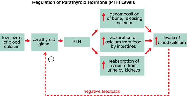
A diagram similar to “Figure 13.18” on page 449 of your textbook would also be acceptable.
SC 20. Emily’s feedback loop would be different because high levels of parathyroid hormone would not inhibit the bone cells from releasing calcium, the kidneys from reabsorbing the calcium, or the intestines from absorbing the calcium from food. All three actions would continue. A feedback loop may resemble the following sample.

SC 21. C
SC 22. D
SC 23. A
SC 24. D
SC 25. C
SC 26. D
SC 27. C
SC 28. A
SC 29. A
SC 30. B
SC 31. 1,7,3,5 (in this order)
SC 32. 4,5,2,4 (in this order)
1.31. Page 5
Module 2—The Endocrine System
 Reflect and Connect
Many articles have been written about the fact that North Americans, including Canadians, consume too much table salt. (Did you know that McDonald’s salts its fries differently in Canada than in the United States?)
Your friend has told you he thinks he consumes way too much table salt, and he has decided to begin a sodium-free diet. He has eliminated table salt, preserved meats, seafood, and dairy products from his diet. Recently, his neck area has become swollen. He feels very tired, is lacking energy, and feels cold all the time even though everyone around him finds the temperature comfortable. He is also astonished to see that he gained weight even though he has little appetite!
Think about what you have learned in this lesson. Based on your friend’s symptoms, what do you think has happened to his endocrine system? Why would you advise him to stop his self-prescribed diet and see a doctor immediately? Why is it important to thoroughly research ideas and consult with your family doctor before beginning a diet such as the one outlined above? You may want to discuss these ideas with your friends and teacher.
Before you begin the Lesson 4 Assignment, you may choose to view all of “The Thyroid Gland: Waging the Battle for Metabolism.”
You may choose to try the questions on page 450 of the textbook. This is an opportunity to discuss any of the concepts in this lesson with your teacher before you begin the Lesson 4 Assignment.
 Module 2: Lesson 4 Assignment
Retrieve the copy of the Module 2: Lesson 4 Assignment that you saved to your computer earlier in this lesson. Complete all questions. Save your completed assignment in your course folder. You will receive instructions about when to submit your assignment to your teacher later in this lesson.
Reflect on the Big Picture
As you have seen, FMEN 1 has far-reaching effects on the endocrine system. The presence of the tiny tumours in Emily’s anterior pituitary, adrenal glands, and parathyroid glands has either led to an undersecretion or an oversecretion of a multiple number of hormones. Loss of homeostasis occurs because chemical communication through negative feedback mechanisms is disrupted.
Using as many of the new terms that you have learned as you can, start drawing a rough draft of a concept map that connects how the anterior pituitary tumours have affected ACTH, TSH, and hGH and their regulatory pathways concerning cortisol, aldosterone, thyroxine, and hGH production. Include how the tumours in the adrenal gland and parathyroid glands have affected epinephrine and PTH production. Use terms such as hypothyroidism, hyperthyroidism, myxedema, cretinism, Grave’s disease, goitre, and iodine. This draft will become part of your final concept map at the end of the module.
Module 2: Lesson 4 Assignment
Submit your completed Module 2: Lesson 4 Assignment to your teacher for assessment.
1.32. Page 6
Module 2—The Endocrine System
 Lesson Summary
This lesson explored the following focusing question:
- How do the thyroid and parathyroid glands contribute to homeostasis?
The thyroid gland, a butterfly-shaped gland located just below the larynx, has embedded in it the four pea-sized parathyroid glands. These glands produce two antagonistic hormones, calcitonin and PTH, that work together to maintain blood calcium homeostasis. Through mechanisms such as increasing absorption of calcium from food or urine and by stimulating decomposition of bone tissue, PTH increases blood calcium levels. Calcitonin decreases blood calcium levels by stimulating uptake of calcium by the bones and inhibiting decomposition of bone tissue. Both are regulated by negative feedback mechanisms.
When stimulated by TSH from the anterior pituitary, the thyroid gland also produces thyroxine, which increases metabolism (cellular respiration) in every cell of your body—especially in cardiac cells, muscle cells, and cells of the kidney and liver. It stimulates the liver to convert glycogen to glucose and increases body temperature. Thyroxine also stimulates proper development and function of the nervous system. Iodine is required to synthesize thyroxine. Lack of thyroxine is called hypothyroidism, which presents differently in babies or children and adults. Overproduction is called hyperthyroidism. Goitre, myxedema, and cretinism are disorders caused by imbalances in thyroxine. Thyroxine levels are regulated by negative feedback of the tropic hormone TSH, which is produced by the anterior pituitary. Surgical removal of parts of the thyroid, death of the most active follicle cells initiated by radioactive iodine, and consumption of thyroid pills are medical technologies that can treat thyroxine imbalances.
Following is a summary of conditions related to the thyroid and parathyroid glands:
- goitre: sometimes called endemic goitre, is an enlargement of the thyroid gland caused by a lack of iodine, which is necessary for the production of thyroxine
- exothalmic goitre: an enlargement of the thyroid gland caused by the overactivity of the thyroid gland due to overstimulation
- hypothyroidism: a disorder of the thyroid gland indicated by a lack of thyroxine, which may be caused by a lack of dietary iodine or a lack of TSH
- hyperthyroidism: overactivity of the thyroid gland that leads to high levels of thyroxine and subsequent speeding up of bodily functions
- myxedema: form of hypothyroidism in adults
- cretinism: a form of hypothyroidism in infants and young children
- Grave’s disease: an autoimmune disorder caused by an abnormal protein that stimulates the thyroid gland to produce and secrete excess thyroxine; the most common cause of hyperthyroidism
Lesson Glossary
Consult the glossary in the textbook for other definitions that you may need to complete your work.
calcitonin: a hormone produced by the C cells (parafollicular cells) in the thyroid gland that decreases blood calcium levels by increasing uptake of calcium into the skeleton and inhibiting decomposition of bone when the blood levels of calcium are too high
C cells (parafollicular cells): cells located between the follicles of the thyroid gland that are responsible for synthesizing calcitonin
cellular respiration: a process occurring in the mitochondria of cells of all living organisms in which energy-rich compounds, such as carbohydrates (glucose), proteins, and fats, are burned with oxygen to release energy in the form of ATP and waste products, such as water and carbon dioxide
follicle: as it relates to the thyroid gland, a microscopic structure consisting of a circle of cells called follicle cells surrounding a central cavity
The thyroid gland consists of many follicles.
metabolism: all of the chemical reactions that occur in living organisms to support and sustain life processes
One type involves the buildup or synthesis of complex compounds from simple ones. A second type consists of the breakdown or decomposition of complex compounds into simple ones. Cellular respiration is one of the major metabolic processes.
precursor: an inactive form of a molecule that can easily be changed into the active form
thyroxine: a hormone produced by the follicle cells of the thyroid gland; mainly controls the rate at which the body metabolizes carbohydrates, fats, and proteins for energy and stimulates the proper development of the nervous system
The precursor thyroglobin can easily be changed into the active thyroxine by the addition of iodine atoms.
1.33. Lesson 5
Module 2—The Endocrine System
Lesson 5—the Pancreas
 Get Focused

left: © Marc Dietrich/shutterstock; right: © Hannamariah/Barbara Helgason/shutterstock
It’s nearly lunchtime. You’re hungry, tired, light-headed, and worried about a big exam in the afternoon. This is stress! What do you grab to eat? Are you the quick candy bar type, or are you the protein-based meat sandwich type? Or will you just skip lunch? Which choice might help you get rid of the symptoms you’re experiencing and get you through the exam without suffering a complete collapse and meltdown?
This situation is all about the pancreas and how it manages high or low blood sugar levels. For a diabetic, this is a daily balancing act—one that can have very serious consequences. Emily’s pancreas cannot regulate her blood sugar level. The tiny tumours growing in the islet cells of her pancreas cause havoc with the levels of her pancreatic hormones. For Emily, the meat sandwich might not make any difference, but the candy bar may save her life.
To understand the role of the pancreas in the endocrine system, you will investigate the following focusing question:
- How does the pancreas contribute to homeostasis?
 Module 2: Lesson 5 Assignment
Your teacher-marked assignment requires that you complete questions about the pancreas and homeostasis.
Download a copy of the Module 2: Lesson 5 Assignment to your computer now. You will receive further instructions about how to complete this assignment later in the lesson.
You must decide what to do with the questions that are not marked by the teacher.
Remember that these questions provide you with the practice and feedback that you need to successfully complete this course. You should respond to all of the questions and place those answers in your course folder.
While you are completing this lesson, there will be many opportunities for you to acquire, understand, and practise the concepts that are presented to you. As you complete these activities, as well as your summary notes, you will file everything in your course folder to reference when you are preparing for exams.
Remember you also have the option of trying additional questions from the textbook for further practice. Consult with your teacher for the answers to these questions. The Key will also provide you with many Diploma Exam-style multiple-choice, numerical-response, and written-response questions that will be an excellent review of the module. Practising your responses to these types of questions is good preparation for the Diploma Exam.
1.34. Page 2
Module 2—The Endocrine System
 Explore
The Anatomy of the Pancreas
pancreas: a gland with dual functions
The non-endocrine part secretes digestive enzymes into the intestine through the pancreatic duct, while the endocrine portion, called the islets of Langerhans, secretes insulin and glucagon into the bloodstream.
islets of Langerhans (islets): clusters of alpha and beta cells that secrete glucagon and insulin, respectively, into the blood
beta cells: cells in the islets that secrete insulin
insulin: a hormone secreted by the beta cells in the islets of the pancreas that lowers blood glucose levels by promoting the uptake of glucose by most cells of the body, and the synthesis and storage of glycogen in the liver; also stimulates protein and fat synthesis
alpha cells: cells in the islets that secrete glucagon
glucagon: a hormone secreted by the alpha cells in the islets of the pancreas that raises blood glucose levels by stimulating liver cells to convert glycogen to glucose; also stimulates fat cells (adipose cells) to convert fat to glucose
The pancreas is a dual-function organ located in the space between the stomach and the first loop of the small intestine. In Biology 20 you learned about the digestive enzymes it produces. Scattered among the enzyme-producing cells are clusters of cells called the islets of Langerhans (or simply islets), which produce pancreatic hormones. The vast majority of cells in the islets are beta cells, which produce insulin. A few elongated alpha cells produce glucagon. Examine the magnified section of “Figure13.24(B)” on page 456 of your textbook to locate these types of cells. (Note: The labels for the alpha and beta cells should be reversed in “Figure 13.24(B).” The beta cells are more numerous.)
Depending on your learning style, you may choose to complete the Read activity or the Watch and Listen activity that follows it.
 Read
Read
To give you a better understanding of the structures of the pancreas, read from the start of page 456 up to the heading “The Effects of Glucose Imbalance” on page 457 in your textbook.
 Watch and Listen
View the segment called “The Pancreas” from “The Pancreas: Regulating Blood Glucose Levels.” You may be required to enter a username and password to access the video. Contact your teacher for this information.
 Self-Check
You may choose to complete SC 1 or SC 2.
SC 1. To apply your knowledge of the anatomy of the pancreas and the function of its various structures, label the structures on the diagram of pancreatic tissue and associated structures. After you have labelled each structure, annotate the structures with their functions. Several structures are shown that you have studied in previous lessons. After you have completed the exercise, check your answers and file your work in your course folder.
Self-Check Answers
SC 1.
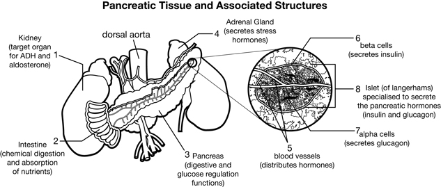
Inquiry into Biology (Whitby, ON: McGraw-Hill Ryerson, 2007), BLM 13.4.1. Reproduced by permission.
SC 2. Practise identifying and labelling the structures of the pancreas by completing this drag-and-drop exercise.
1.35. Page 3
Module 2—The Endocrine System
Pancreatic Hormones
The study of the pancreatic hormones is a significant section of this module. Calcitonin and PTH, which are produced by the thyroid and parathyroid glands, work against each other to control the level of calcium in the blood. Insulin and glucagon, which are produced by the pancreas, are another pair of antagonistic hormones. They work against each other to regulate blood glucose level. Insulin lowers blood glucose levels, whereas glucagon raises blood glucose levels. Together, these hormones maintain homeostasis by way of negative feedback. Restoration to a range of normal glucose levels acts to control hormone secretion.
You may choose to do either the Read activity or the Watch and Listen activity to explore how pancreatic hormones maintain blood glucose levels. If you wish, do both.
 Read
Read
Review pages 456 and 457 of your textbook, which describe how pancreatic hormones maintain blood glucose levels. As you read, make notes. A good summary of negative feedback is given in “Figure 13.25” on page 457 of the textbook. Store your notes, including the feedback loop diagram, in your course folder.
 Watch and Listen
Watch the following two segments of “The Pancreas: Regulating Blood Glucose Levels.” Make relevant notes to be stored for later use. You may be required to enter a username and password to access these videos. Contact your teacher for this information.
- “Blood Glucose Levels”
- “Bio Review: Blood Glucose Levels”
 Try This
It is important to understand the role of insulin and glucagon in the regulation of glucose levels in the blood. To check your understanding, complete the following closed written-response question that has four parts. This format is similar to the closed written-response questions on the Diploma Exam. Notice that mark values have been assigned for each part. The key to this exercise will explain how the mark values are assigned. When you check your answers, mark each part accordingly. These mark values should help you develop comprehensive answers as required on the Diploma Exam. After checking this exercise, file it in your course folder. Consult your teacher if you require clarification or help.
Use the following information to answer question TR 1.
Blood glucose concentrations of one individual were monitored over several hours. The following data was collected. The blood glucose level of a healthy person is approximately 90 mg/100 mL.
|
TR 1. Plot this data on a grid like the one provided below. Draw a curved line through the points and label your graph appropriately. Draw a horizontal dotted line showing the approximate blood glucose level of a healthy individual. (6 marks)

TR 2. On the graph, indicate with an arrow two places where a blood test would reveal high levels of insulin. Explain why insulin would be at high levels at these times. (4 marks)
TR 3. Predict what would happen to this person’s blood glucose levels if he or she worked out at the gym from 9:00 a.m. to 10:00 a.m. With respect to blood glucose levels, how would this person’s body respond? (3 marks)
TR 4. The graph shows that this person’s glucose levels fluctuate throughout the day. Is this person healthy or does he or she have a malfunctioning pancreas? Explain your answer. (3 marks)
 Try This
TR 1. Your graph should look similar to the one shown below.
Marking Guide
1 mark for the title, which must include the manipulated and responding variable
1 mark for correct labels and units on the x-axis
1 mark for correct labels and units on the y-axis
1 mark for using suitable scales for each axis (graph should use the whole grid)
1 mark for drawing a smooth, curved line through the points rather than a jagged line
1 mark for showing the approximate blood glucose level of a healthy individual
TR 2. The first place where insulin levels would be high would be a time somewhere between 7:00 a.m. and 8:00 a.m. This should be marked on the graph with an arrow. (1 mark) This is a time shortly after breakfast. The absorption of glucose from the meal would raise blood glucose levels, and the high glucose levels would stimulate the beta cells in the pancreas to release insulin to lower blood glucose levels back to the normal range. (1 mark for the explanation)
The second place that insulin would be high would be a time somewhere between 12:00 noon and 1:00 p.m. This should be marked on the graph with an arrow. (1 mark) This is a time shortly after lunch. Absorption of glucose from the meal would raise blood glucose levels, and the high glucose levels would stimulate the beta cells in the pancreas to release insulin to lower blood glucose levels back to the normal range. (1 mark for explanation)
TR 3. The muscle cells would use increased amounts of glucose for cellular respiration. (1 mark) As a result, blood glucose levels would start to drop. (1 mark) In response to lower blood glucose levels, the alpha cells in the pancreas would secrete glucagon into the bloodstream. Glucagon stimulates the liver to convert glycogen into glucose, which would be released into the blood to raise blood glucose levels. (1 mark)
TR 4. This individual is healthy according to the levels of blood glucose indicated. (1 mark) If 90 mg/100 mL is considered to be a normal set point, then small fluctuations around the set value are to be expected. (1 mark) It is only if the fluctuations are large and stay above or below the set point continually that the person is considered as having pancreatic dysfunction. (1 mark)
1.36. Page 4
Module 2—The Endocrine System

© PeJo/shutterstock
Blood Glucose Imbalances
Among your friends, relatives, and teachers, you are likely to know someone who has diabetes mellitus. This is a disorder in which blood glucose levels are abnormally high. Doctors usually use the full name rather than just “diabetes” to distinguish it from diabetes insipidus. In Biology 20 you learned about diabetes insipidus. This disorder is also hormone related, but it involves insufficient secretion of antidiurectic hormone (ADH). Many of the symptoms, however, are similar to diabetes mellitus, such as excessive thirst, weakness, and frequent urination.
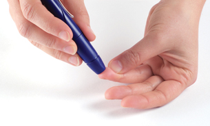
© Adam Majchrzak/shutterstock
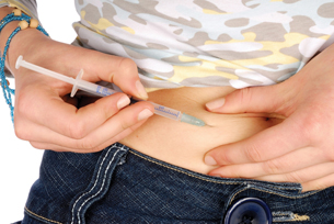
© ajt/shutterstock
diabetes mellitus: a serious chronic disorder that results when the pancreas does not produce enough insulin, insulin receptors, or body cells do not respond to insulin
Levels of blood glucose tend to rise sharply (spike) after meals (hyperglycemia) and remain at significantly elevated levels.
hyperglycemia: a condition resulting from high levels of blood glucose; occurs in individuals with diabetes mellitus
hypoglycemia: a condition resulting from low levels of blood glucose; occurs in individuals who secrete excessive amounts of insulin when a tumour develops in the beta cells or in diabetics who have injected too much insulin
Type 1 diabetes(juvenile diabetes or insulin-dependent diabetes): an autoimmune disorder in which the immune system produces antibodies that attack and destroy the beta cells of the pancreas so that they are unable to produce insulin; usually diagnosed at an early age and the individuals require daily injections of insulin
Type 2 diabetes (adult-onset diabetes or insulin-independent diabetes): a disorder that develops slowly over time because the insulin receptors on the body’s cells stop responding to insulin or because the beta cells of the pancreas produce less and less insulin over time; appears to be related to being overweight or obese
Diabetes is usually grouped as Type 1 diabetes (also called insulin dependent or juvenile diabetes). In Type 1 diabetes, the pancreas does not produce insulin. In Type 2 diabetes (also called insulin-independent or adult-onset diabetes), the body may reduce its production of insulin or the body’s insulin receptor cells may no longer be sensitive to insulin.
Lack of insulin causes blood glucose levels to spike after meals and to remain high for long periods of time. This is called hyperglycemia. If there is too much insulin, glucose levels plummet, and the person has hypoglycemia (a hypoglycemic episode).
Untreated glucose imbalances lead to many symptoms, and can result in blindness, kidney failure, nerve damage, and severe infection in the limbs, which may lead to the need for amputation. Several medical technologies have been developed to help diabetics deal with their problems.
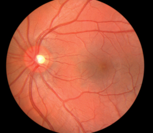
© Tim Mainiero/iStockphoto
Depending on your learning style, you may choose to complete the following Read activity or the Watch and Listen activity.
 Read
Read
Read “The Effects of Glucose Imbalance” on pages 457 through 459 of your textbook. As you read, make notes about the types of diabetes, their causes, their symptoms, their effects, and the medical technologies that have been developed to deal with glucose imbalances. File your work in your course folder.
 Watch and Listen
View the following segments of “The Pancreas: Regulating Blood Glucose Levels.” Make notes about the types of diabetes, their causes, their symptoms, their effects, and the medical technologies that have been developed to deal with glucose imbalances. You may require a username and password to access these videos. Contact your teacher for this information.
- “Type 1 Diabetes”
- “Bio Fact: Diabetes Mellitus”
- “Urinalysis”
- “Bio STS: Blood Glucose Monitor”
- “Bio Quest: Research Approaches for Diabetes Mellitus”
- “Working with Diabetes”
1.37. Page 5
Module 2—The Endocrine System
 Self-Check
Apply what you have learned about diabetes mellitus by completing the following questions. Check your answers and file your work. Consult your teacher for clarification of any concepts you may not understand.
SC 3. Complete the flow chart that illustrates normal blood glucose regulation through negative feedback.
SC 4. Complete a table like the one shown by comparing Type 1 diabetes with Type 2 diabetes.
A Comparison of Type 1 Diabetes with Type 2 Diabetes
| Feature | Type 1 Diabetes | Type 2 Diabetes |
|---|---|---|
| Cause | ||
| Onset | ||
| Age of Onset | ||
| Predisposition | ||
| Insulin Production | ||
| Immediate Symptoms | ||
| Long Term Effects | ||
| Treatment |
SC 5. FMEN 1 wreaks havoc with the levels of Emily’s pancreatic hormones. At different times in her life, tumours have grown in different areas of her pancreas. When tumours affect the beta cells, there is an oversecretion of insulin. Emily then becomes hypoglycemic.
- Differentiate between hyperglycemia and hypoglycemia.
- Describe what happens to the blood glucose level when there is an oversecretion of insulin.
- Outline how a person, such as Emily, reacts to hypoglycemia.
- Explain what other hormone may also be oversecreted as the pancreas tries to deal with the low blood glucose levels.
- What can Emily do to alleviate the symptoms quickly?
Use the following graph to answer question SC 6.
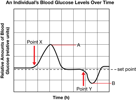
SC 6.
- What occurred at Point X?
- Identify the endocrine secretion represented by A.
- What happened at Point Y?
- Identify the endocrine secretion represented by B.
Self-Check Answers
SC 3. The completed flow chart representing normal regulation of blood glucose through negative feedback should resemble the following.

SC 4. The table comparing Type 1 and Type 2 diabetes should resemble the following.
A Comparison of Type 1 Diabetes with Type 2 Diabetes
| Feature | Type 1 Diabetes | Type 2 Diabetes |
|---|---|---|
| Cause | autoimmune disease where antibodies destroy beta cells in pancreas and little or no insulin is produced |
receptors on target cells do not respond to the insulin produced; insulin resistance develops |
| Onset | rapid |
gradual |
| Age of Onset | childhood |
adulthood, although more cases in young people recently |
| Predisposition | none known |
obesity increases risk of development significantly |
| Insulin Production | very little or none |
insulin produced but ineffective |
| Immediate Symptoms | fatigue, low energy, glucose in urine, increased volume of urine, increased thirst, weight loss, smell of nail polish on breath |
fatigue, low energy, glucose in urine, increased volume of urine, increased thirst, no smell of nail polish on breathe |
| Long Term Effects | blindness, kidney failure, nerve damage, gangrene, increased risk of heart attack and stroke |
blindness, kidney failure, nerve damage, gangrene, increased risk of heart attack and stroke |
| Treatment | insulin injections, islet transplantation in experimental stages |
control diet, lose weight, increase exercise, drugs to decrease insulin resistance |
SC 5.
- Hyperglycemia occurs when blood glucose levels rise significantly over normal values and stay at an elevated level. This condition is usually caused by a lack of insulin. Hypoglycemia occurs when blood glucose levels fall significantly below normal values and are not raised. This condition is caused by the oversecretion of insulin.
- When there is an oversecretion of insulin, cells are stimulated to take up large amounts of glucose and the liver and muscle cells are stimulated to convert large amounts of glucose to glycogen for storage. Adipose (fat) cells are stimulated to convert glucose to fat. All actions significantly lower blood glucose levels below normal values.
- When a person, such as Emily, is hypoglycemic, he or she becomes anxious, nervous, develops trembling in his or her hands, and has a feeling of weakness. Insufficient glucose to the brain cells causes disorientation, which may progress to convulsions, coma, and possibly death.
- Through negative feedback, low blood glucose levels stimulate the alpha cells in the pancreas to secrete glucagon, which instructs cells to convert glycogen back to glucose as well as to convert non-carbohydrates such as fats and proteins to glucose.
- Emily can raise her blood glucose levels by eating something with a lot of glucose in it or she can inject glucagon.
SC 6.
- At Point X, the person ate something with sugar in it.
- A represents the secretion of insulin by the pancreas.
- At Point Y, the person probably did some exercise.
- B represents the secretion of glucagon by the pancreas.
1.38. Page 6
Module 2—The Endocrine System
 Self-Check
Apply your understanding of the concepts on the pancreas and its regulation of blood glucose by answering the multiple-choice and numerical-response questions that follow. Check your answers. If you do not understand any questions, consult your teacher.
Multiple-Choice Questions
SC 7. Which of the following rows correctly matches the endocrine gland to the hormone it secreted and to the effect caused by the hormone?
Row |
Endocrine Gland |
Hormone Secreted |
Target Cell/Organ |
A. |
thyroid gland |
thyroxine |
increases blood glucose |
B. |
pancreas |
insulin |
increases blood glucose |
C. |
pancreas |
glucagon |
increases blood glucose |
D. |
adrenal cortex |
epinephrine |
increases blood glucose |
SC 8. Which hormone is not correctly matched to the disorder or disease associated with it?
- insulin and diabetes mellitus
- aldosterone and diabetes insipidus
- thyroxine and goitre
- cortisol and depressed immune system
SC 9. Which of the following hormones does not affect the carbohydrate metabolism?
- human growth hormone
- epinephrine
- insulin
- ADH
SC 10. Which of the following endocrine glands does not produce a hormone that directly affects blood glucose levels?
- posterior pituitary
- pancreas
- adrenal glands
- thyroid gland
SC 11. Which of the following explains one of the differences between Type 1 and Type 2 diabetes?
- The treatment for Type 2 diabetes involves insulin injections, while Type 1 can usually be controlled by diet.
- Only Type 1 can result in complications such as kidney disease, reduced circulation, or stroke.
- Type 1 can be a result of lifestyle, and Type 2 is thought to be caused by a virus or other agent.
- People with Type 2 diabetes can produce insulin, but this insulin cannot be used. Type 1 diabetes results from lack of insulin production.
SC 12. Which pair consists of antagonistic hormones?
- thyroxine and calcitonin
- epinephrine and norepinephrine
- cortisol and epinephrine
- insulin and glucagon

SC 13. Which of the following rows correctly matches the endocrine gland labelled in the diagram to the hormone it produces?
Row |
Label on Diagram |
Endocrine Gland |
Hormone |
A. |
E |
adrenal gland |
cortisol and thyroxine |
B. |
A |
pituitary gland |
releasing hormone and ADH |
C. |
F |
pancreas |
insulin and glucagon |
D. |
C |
thyroid |
TSH and thyroxine |
SC 14. Which endocrine gland shown on this diagram would be directly responsible for the development of diabetes mellitus in humans?
- B
- D
- E
- F
SC 15. Symptoms resulting from low secretions of the hormones produced by the gland labelled F include
- high levels of sodium and sugar in the urine
- increased thirst and decreased urine production
- loss of weight and high levels of glucose in the urine
- obesity and increased risk of stroke
SC 16. The endocrine function of the pancreas was studied in Canada using dogs as experimental animals. The pancreatic cells with an endocrine function are
- islet cells
- C cells
- follicle cells
- interstitial cells
Use the following diagram to answer question SC 17.
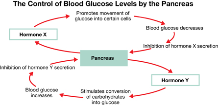
SC 17. Hormones X and Y, respectively, are
- insulin and glucagon
- glucagon and insulin
- insulin and epinephrine
- epinephrine and insulin
Use the following information to answer questions SC 18 and SC 19.
A Treatment for Pancreatitis
Surgical removal of the pancreas is a procedure that doctors use to relieve the pain of patients suffering from chronic pancreatitis (inflammation of the pancreas). Unfortunately, the surgery causes the onset of diabetes mellitus and other complications in these patients. In a recent study involving five individuals who had undergone this treatment, islet cells from the removed pancreas were infused (transplanted) back into the liver of each patient. This procedure effectively eliminated the occurrence of diabetes in these patients. |
SC 18. Blood glucose levels are kept relatively constant by a negative feedback mechanism. The islet cells are part of this mechanism. The role of these cells is to secrete
- glucagon to raise blood glucose levels, and insulin to lower blood glucose levels
- glucagon to lower blood glucose levels, and insulin to raise blood glucose levels
- glucagon and insulin, both of which lower blood glucose levels
- glucagon and insulin, both of which raise blood glucose levels
SC 19. Patients who participated in this study no longer have a pancreas. To maintain normal body functions in these patients, the infusion of islet cells would have to be accompanied by a daily intake of
- glycogen to stimulate liver function
- hormones to promote glycogen release from the liver
- digestive enzymes into the blood to maintain nutrient levels
- digestive enzymes to replace those produced by the pancreas
Numerical-Response Questions
Use the following information to answer question SC 20.
Responses Stimulated by Hormones
|
SC 20. Identify the response, as numbered above, that would be stimulated by each of the hormones given below.
Response:
______ ______ ______ ______
Hormone: Insulin Cortisol Glucagon hGH
Use the following information to answer question SC 21.
Some Endocrine Glands and Hormones
|
SC 21. To complete this statement, select the number of the gland or hormone from the information above that best completes each blank.
The hormones of the _____ act antagonistically to regulate blood glucose levels. The beta cells of the islets of Langerhans secrete ____, which lowers blood glucose levels. The alpha cells secrete _____, which raises blood glucose levels. Type 1 diabetes causes hyperglycemia because the secreting cells have degenerated. Type 2 diabetes tends to develop gradually, often because the _____ receptors on the body’s cells stop responding to this hormone.
Self-Check Answers
SC 7. C
SC 8. B
SC 9. D
SC 10. A
SC 11. D
SC 12. D
SC 13. C
SC 14. D
SC 15. C
SC 16. A
SC 17. A
SC 18. A
SC 19. D
SC 20. 5, 4, 6, 2 (in that order)
SC 21. 4, 2, 5, 2 (in that order)
1.39. Page 7
Module 2—The Endocrine System
 Reflect and Connect
Suppose you have a 92-year-old grandfather who lives in an extended care facility. He is sharp as a tack, but has Type 2 diabetes mellitus. He is very conscientious about making sure that he takes his medication regularly. You go to visit him, and he tells you about an incident that really scared him. He relates how he had become ill and experienced vomiting and diarrhea all day. He thinks that this was caused by the institutional food! He lost his appetite for eating but still tried to take his medication. A nurse found him unconscious on the floor of his room. Luckily, the facility physician diagnosed the cause and helped him. He asks you to help him understand what happened. What would you say?
Think about what you have learned in this lesson. Can you suggest what condition may have occurred in your grandfather’s blood because he stopped eating but tried to continue his medication? Can you predict what the physician did to treat your grandfather? Did your grandfather forget to monitor his blood glucose? Should he have been taking the medication if he was not eating? Did he know this? What information would you review with your grandfather about the role of his medicine? You may want to discuss these questions with your teacher to verify your ideas. Then, proceed with the Lesson 5 Assignment.
Before you begin the Lesson 5 Assignment, ask your teacher which questions on page 462 of the textbook you should use as a review. You may also choose to watch the entire “The Pancreas: Regulating Blood Glucose Levels” video. If you have any questions, consult with your teacher.
 Module 2: Lesson 5 Assignment
Retrieve the copy of the Module 2: Lesson 5 Assignment that you saved to your computer earlier in this lesson. Complete all questions. Save your completed assignment in your course folder. You will receive instructions about when to submit your assignment to your teacher later in this lesson.
Reflect on the Big Picture
Think about your case study of Emily. The tumours caused by FMEN 1 usually caused excessive secretion of various hormones. The exception was the undersecretion of thyroid stimulating hormone by the anterior pituitary.
As you review this lesson, apply your knowledge to develop hypotheses about the following items:
- the effect the tumours in the islets of the pancreas had on Emily
- the effect to Emily’s blood glucose levels
- whether or not Emily became a diabetic
- technologies and treatments that would be available, and how they would help Emily establish homeostasis
 Going Beyond
A University of Alberta research team has developed a procedure called the Edmonton Protocol that helps people who have difficulty managing Type 1 diabetes undergo an islet transplant procedure. On page 466 in your textbook, read the interview with Dr. Edmond Ryan, Medical Director of the University of Alberta Clinical Islet Transplant Program.
- Research how islet transplantation in the Edmonton Protocol is performed.
- Islet transplant patients must wait for the treatment until donated organs become available. Outline three other potential sources of islet cells and outline the issues related with each source.
- Explain why stem cell research is important to islet cell transplantation.
Module 2: Lesson 5 Assignment
Submit your completed Module 2: Lesson 5 Assignment to your teacher for assessment.
1.40. Page 8
Module 2—The Endocrine System
 Lesson Summary
© Lisa A. Svara/shutterstock
Communication of information to and from a command centre in a battle is essential for success. The pancreas is like a command centre that monitors blood glucose. High levels of glucose stimulate the beta cells in the islets of the pancreas to secrete insulin, which increases the passage of glucose into body cells and promotes the conversion of glucose to glycogen in liver and muscle cells and to fat in the adipose cells. Low glucose levels stimulate the pancreatic alpha cells in the islets to secrete glucagon, which promotes the breakdown of glycogen to glucose and the release of this glucose into the bloodstream.
When the communication is disrupted, the battle may be lost. So it is with blood glucose regulation. If the beta cells are destroyed by an autoimmune disease, and little or no insulin is produced, consistently high blood glucose levels result in Type 1 diabetes. Left untreated, life-threatening consequences result.
© Daniel Gale/shutterstock
Regular glucose monitoring and insulin injections are the only treatment. Insulin cannot be taken in pill form because the digestive system would break it down before it could be absorbed.
If the pancreas sends out insulin, but the body cells ignore it (called insulin resistance), Type 2 diabetes results. Being very fat (obese) increases the cells inability to respond to insulin. The obvious treatment is to lose weight. Controlling diet and regular exercise can persuade the cells to respond to the insulin. In some cases, drugs have to be used.
Sometimes the pancreas secretes too much insulin and hypoglycemia occurs. The person becomes anxious or nervous, develops shakiness, and has a feeling of weakness. Continued hypoglycemia causes the brain to become disoriented, and may progress to unconsciousness and possibly death. If symptoms are recognized quickly, it is easily treated by eating candy, a source of glucose, or injecting glucagon.
Medical technologies for dealing with diabetes mellitus have progressed from glucose dipsticks to digital glucose monitors, and from syringes to insulin pens to insulin pumps. Research is being done into treating diabetes mellitus with islet transplants.
Lesson Glossary
Consult the glossary in the textbook for other definitions that you may need to complete your work.
alpha cells: cells in the islets that secrete glucagon
beta cells: cells in the islets that secrete insulin
diabetes mellitus: a serious chronic disorder that results when the pancreas does not produce enough insulin, insulin receptors, or body cells do not respond to insulin
Levels of blood glucose tend to rise sharply (spike) after meals (hyperglycemia) and remain at significantly elevated levels.
glucagon: a hormone secreted by the alpha cells in the islets of the pancreas that raises blood glucose levels by stimulating liver cells to convert glycogen to glucose; also stimulates fat cells (adipose cells) to convert fat to glucose
hyperglycemia: a condition resulting from high levels of blood glucose; occurs in individuals with diabetes mellitus
hypoglycemia: a condition resulting from low levels of blood glucose; occurs in individuals who secrete excessive amounts of insulin when a tumour develops in the beta cells or in diabetics who have injected too much insulin
insulin: a hormone secreted by the beta cells in the islets of the pancreas that lowers blood glucose levels by promoting the uptake of glucose by most cells of the body, and the synthesis and storage of glycogen in the liver; also stimulates protein and fat synthesis
islets of Langerhans (islets): clusters of alpha and beta cells that secrete glucagon and insulin, respectively, into the blood
pancreas: a gland with dual functions
The non-endocrine part secretes digestive enzymes into the intestine through the pancreatic duct, while the endocrine portion, called the islets of Langerhans, secretes insulin and glucagon into the bloodstream.
Type 1 diabetes (juvenile diabetes or insulin-dependent diabetes): an autoimmune disorder in which the immune system produces antibodies that attack and destroy the beta cells of the pancreas so that they are unable to produce insulin; usually diagnosed at an early age and the individuals require daily injections of insulin
Type 2 diabetes (adult-onset diabetes or insulin-independent diabetes): a disorder that develops slowly over time because the insulin receptors on the body’s cells stop responding to insulin or because the beta cells of the pancreas produce less and less insulin over time; appears to be related to being overweight or obese
1.41. Lesson 6
Module 2—The Endocrine System
Lesson 6—Bringing It Together
 Get Focused
In the previous module you learned how the nervous system responds to environmental stimuli by using neural pathways to produce very rapid and precise responses. The endocrine system responds to stimuli by using chemical messengers in the blood to control metabolic processes in the body. These responses are much slower than the responses of the nervous system, with the exception of the responses controlled by epinephrine. Your body is in balance (homeostasis) because your nervous system responds to immediate stimuli or crises while your hormonal system maintains long-term stability. A major part of this balance is missing from Emily’s life. Because of the tumours that can grow on any of her glands, her endocrine system does not respond appropriately. The balance of Emily’s endocrine system is jeopardized, impacting her health and life. The decisions you make, and the overall health of your nervous and endocrine systems, can impact your life now and in the long run.
This lesson will allow you to explore how the nervous and endocrine systems complement and communicate with each other to maintain homeostasis and well-being.
In this lesson you will investigate the following focusing question:
- How are the nervous and endocrine systems interdependent and how are they different?
 Module 2: Lesson 6 Assignment
Download a copy of the Module 2: Lesson 6 Assignment to your computer now. You will receive further instructions about how to complete this assignment later in the lesson.
You must decide what to do with the questions that are not marked by the teacher.
Remember that these questions provide you with the practice and feedback that you need to successfully complete this course. You should respond to all of the questions and place those answers in your course folder.
Because this is the last lesson in this module, you will also need to submit the Module Assessment, which is a written-response question. The question is based on data from a case study involving one of the hormones that you have studied in this module. When you have completed the module, go to the Module Summary and Assessment page for information about the written-response question and the evaluation criteria.
While you are completing this lesson, there will be many opportunities for you to acquire, understand, and practise the concepts that are presented to you. As you complete these activities, as well as your summary notes, you will file everything in your course folder to reference when you are preparing for exams.
Remember you also have the option of trying additional questions from the textbook for further practice. Consult with your teacher for the answers to these questions. The Key will also provide you with many Diploma Exam-style multiple-choice, numerical-response, and written-response questions that will be an excellent review of the module. Practising your responses to these types of questions is good preparation for the Diploma Exam.
1.42. Page 2
Module 2—The Endocrine System
 Explore
Regulating the Internal Environment
You and your friends are at a rock concert. Coloured lights are flashing, and the music is loud. You are dancing with your friends and having a great time. You see the changing lights, you hear and feel the music, and you sense the temperature of the room. Heart rate, breathing rate, body temperature, and sweating are all increased.
Your body is able to detect and respond to environmental change and is able to regulate your internal environment by the two control systems you have studied in this unit—the nervous and endocrine systems. Although they are structurally different in many ways, the two systems must constantly interact to control physiological processes such as heart rate.
 Read
Read
Read pages 436 to 437 in your textbook to explore how the nervous and endocrine systems are different.
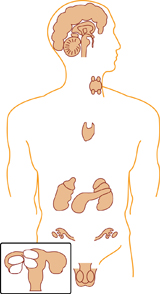
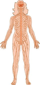
© Oguz Aral/shutterstock
 Self-Check
SC 1. To summarize the differences between the nervous and endocrine systems, complete a table like the following that contrasts the two systems with respect to method of communication, speed of communication, duration of response, target pathway, and what action is brought about by the effectors. Check your answers and file your work in your course folder.
Feature To Be Contrasted |
Nervous System |
Endocrine System |
Method of Communication |
|
|
Speed |
|
|
Duration |
|
|
Target Pathway |
|
|
Action |
|
|
Self-Check Answers
SC 1. Your table should resemble the one below.
Feature To Be Contrasted |
Nervous System |
Endocrine System |
Method of Communication |
neural pathways or electrochemical impulse transmission |
hormones in bloodstream |
Speed |
very rapid (milliseconds) |
relatively slow (minutes/hours/days) |
Duration |
short-term effects |
longer-lasting effects |
Target Pathway |
specific through nerves to glands or muscles |
hormones to a broad range of target cells |
Action |
causes glands to secrete or muscles to contract |
causes changes in metabolic activity |
1.43. Page 3
Module 2—The Endocrine System
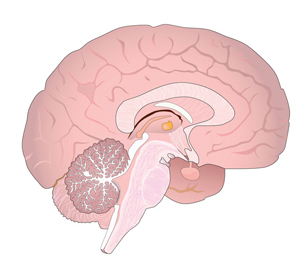
© Cristian Alexandru Ciobanu/shutterstock
The Brain and the Pituitary Gland
The hypothalamus, which is located just above the pituitary gland, is an important part of the brain. The pituitary gland, however, is an important part of the endocrine system. Together, the hypothalamus and the pituitary gland form an important communication link between the nervous system and the endocrine system.
The hypothalamus contributes to the homeostasis of the nervous system. Information from the sense organs is transmitted through sensory pathways to the hypothalamus. Based on this information, the hypothalamus co-ordinates many basic physiological activities, including the reflex activity of the autonomic nervous system.
The hypothalamus also contributes to the homeostasis of the endocrine system. There are essentially three groups of nerve centres in the hypothalamus. These centres allow communication between the nervous and endocrine systems. They send instructions to either the anterior or the posterior lobe of the pituitary. The pituitary, the master gland of the endocrine system, is then stimulated to increase or decrease secretion of its hormones into the bloodstream. The first nerve centre controls the secretion of releasing hormones from the hypothalamus. These releasing hormones are carried through the blood vessels to the anterior pituitary, where they regulate the secretion of hormones, such as thyroid stimulating hormone (TSH), from the anterior pituitary. The second nerve centre performs two functions in its regulation of the pituitary gland:
- It secretes hormones that are carried by the nerve cells to the posterior pituitary, where they are stored in the axon terminals.
- It sends nerve impulses to the posterior pituitary, stimulating the release of the stored hormones, such as antidiuretic hormone (ADH) and oxytocin.
The third centre in the hypothalamus is connected by sympathetic neurons to the adrenal gland, regulating the release of epinephrine.
These examples illustrate the importance of the hypothalamus as an important command centre for co-ordinating the activity of the nervous and endocrine systems.
Depending on your learning style, you may choose to compete the Read activity or the Watch and Listen activity that follows it.
 Read
Read
Review page 437 and page 463 of your textbook, and finalize your notes about how the nervous and endocrine systems maintain homeostasis, how they are co-ordinated, and how they act together. Developing a flow chart may help you visualize these relationships. You may want to review your notes from Lessons 1, 2, 3 and 4 of Module 2 before you begin. Post your notes for discussion with your peers and your teacher. File your notes in your course folder.
 Watch and Listen
Watch the following segments of “The Neuroendocrine System: Working Together to Maintain Homeostasis.” Make notes as you watch. File your notes in your course folder. You may be required to enter a username and password in order to access these videos. Contact your teacher for this information.
- “Nervous and Endocrine System”
- “Bio Bit: Nervous vs. Endocrine Action”
- “Bio Review: Basic Nervous/Endocrine Differences”
- “Blood Glucose Regulation”
 Self-Check
Complete the following questions to support your understanding. Check your answers and then file your work in your course folder. Consult with your teacher if you have questions.
SC 2. Using an example, explain how the nervous and endocrine systems work together to regulate a response in the body.
SC 3.
- Why has the pituitary gland often been called the “master gland”?
- Explain how the hypothalamus controls the pituitary gland.
- Is “master gland” a suitable name for the pituitary gland?

© Enge/iStockphoto
SC 4. Explain how the anterior pituitary and posterior pituitary differ with respect to their relationship to the hypothalamus.
SC 5. How are neurotransmitters and hormones similar? How are they different?
SC 6. Using an example, explain how the hormones of the adrenal medulla complement the actions of the sympathetic branch of the autonomic nervous system.
SC 7. Compare and contrast the role of the norepinephrine in the nervous system with its role in the endocrine system.
SC 8. How is the hypothalamus involved in the release of epinephrine and norepinephrine by the adrenal gland?
SC 9. How is the secretion of tropic hormones from the pituitary gland regulated?
SC 10. Compare the anatomy of the pituitary gland and adrenal medulla.
SC 11. Why is control of the adrenal medulla by the nervous system important?
Self-Check Answers
SC 2. Your answers may contain one of the following:
- Some nervous system tissues secrete hormones. For example, neurosecretory cells in the hypothalamus produce antidiuretic hormone and oxytocin, which are stored and released by the posterior pituitary.
- Several chemicals function as body neurotransmitters and hormones, depending on their location in the body. For example, both epinephrine and norepinephrine act as a neurotransmitters in synaptic transmission in certain neural pathways, such as the sympathetic nervous system, as well as hormones released by the adrenal medulla in the fight-or-flight response (short-term stress).
- The regulation of several physiological processes involves both the nervous and endocrine systems acting in conjunction with each other. When a mother breast feeds her baby, the baby’s suckling initiates sensory messages in the mother’s neurons that travel to the hypothalamus, which in turn triggers the pituitary to release a hormone called oxytocin. Oxytocin travels in the bloodstream to the mammary glands of the breast, causing the secretion of milk. Another example that might be outlined is that of sensory messages of something that causes excessive fear or rage. In such a case, a message is relayed to the hypothalamus which initiates nerve impulses in the sympathetic nervous system, which in turn stimulates secretory cells in the adrenal medulla to secrete epinephrine, which initiates a response to the stress.
SC 3.
- The pituitary gland is often referred to as the “master gland” because it synthesizes many hormones and releases many tropic hormones that regulate the action of other endocrine glands.
- Neurosecretory cells in the hypothalamus produce releasing and release-inhibiting hormones. These hormones are secreted into the blood vessels between the hypothalamus and anterior pituitary. Each type of hormone from the hypothalamus either stimulates or inhibits production and secretion of an anterior pituitary hormone into the bloodstream.
- Your answers could either support or reject the “master gland” label assigned to the pituitary gland. Some may argue that the hypothalamus is part of the brain and not a separate endocrine gland. In this case, the name “master gland” given to the pituitary gland is suitable. Others may argue that the hypothalamus should be called the “master gland” because it is actually controlling the pituitary gland through releasing hormones.
SC 4. The posterior pituitary is an extension of the hypothalamus, and both are composed of specialized nerve cells. Hormones produced in the hypothalamus are transported by nerve cells to the posterior pituitary, where they are stored and released when stimulated by neurons from the hypothalamus. On the other hand, the anterior pituitary is composed of secretory cells that produce and release hormones when stimulated by hormones from the hypothalamus, which travel to the pituitary by way of the bloodstream.
SC 5. The similarities between neurotransmitters and hormones include the following:
- Both are stored in cells for later release.
- Stimulation of cells prompts release.
- Some molecules function as both a hormone and a neurotransmitter.
- Both require a specific receptor for action.
- Both include responses that are regulated by negative feedback loops.
The differences between neurotransmitters and hormones include the following:
- Neurotransmitters act more rapidly than hormones.
- Neurotransmitters are released in synapses between neurons, whereas hormones are secreted into the bloodstream, where they are transported to a broader range of cell types.
SC 6. The sympathetic nervous system and the adrenal medulla produce hormones that regulate a short-term response to stress, commonly referred to as the fight-or-flight response. In response to a stress stimulus, such as fear, neurons of the sympathetic nervous system release a neurotransmitter called norepinephrine. Other neurons from the sympathetic nervous system carry a signal from the hypothalamus directly to the adrenal medulla. These neurons, rather than hormones, stimulate the adrenal medulla to secrete epinephrine or norepinephrine. The combined action of the neurotransmitter and the hormones triggers the fight-or-flight response, which is characterized by an increase in breathing rate, heart rate, blood pressure, blood flow to the heart and muscles, and the conversion of glycogen to glucose in the liver.
SC 7. Norepinephrine is released by neurons of the sympathetic nervous system and by the cells of the adrenal medulla. The sympathetic neurons release norepinephrine as a neurotransmitter for synaptic transmission. In response to a stress stimulus, neurons of the sympathetic nervous system carry a signal from the hypothalamus directly to the adrenal medulla. These neurons, rather than hormones, stimulate the adrenal medulla to secrete norepinephrine or epinephrine. These hormones also trigger the fight-or-flight response. One difference is that the response to danger by the nervous system is much faster than that of the endocrine system. Another difference is that the effects of norepinephrine released by the adrenal medulla last longer.
SC 8. Centres in the hypothalamus initiate an impulse in a sympathetic neural pathway which connects to the secretory cells in the adrenal medulla and stimulates them to release epinephrine or norepinephrine, resulting in the fight-or-flight response.
SC 9. Typically, the hypothalamus secretes a releasing hormone that is transported by the blood to the anterior pituitary gland. This causes the anterior pituitary to release a tropic hormone into the bloodstream. The pituitary tropic hormone then stimulates the target gland to release its hormone into the blood. This third hormone travels to another target tissue and produces an effect. Levels of the third hormone prevent further release of the first two hormones in the pathway by way of negative feedback.
SC 10. The anatomy of the posterior pituitary is similar to that of the adrenal medulla as both are composed of modified nerve cells and release hormones. The anatomy of the anterior pituitary and the adrenal cortex are similar in that both are composed of secretory cells that synthesize hormones.
SC 11. It is important for the adrenal medulla to be stimulated by the nervous system so that responses are very rapid rather than slower, as would occur if it was stimulated by the endocrine system. Fight-or-flight responses are often life-preserving and require quick action.
1.44. Page 4
Module 2—The Endocrine System
 Reflect and Connect
You will now have the opportunity to reflect on the glands and hormones of the endocrine system and how they contribute to homeostasis. You will do this by considering Emily and the consequences of having FMEN 1. Answer the following questions and post your response to the course discussion area. Reflect on and respond to at least two other responses.
Respond to the following questions:
- In Emily’s case, as far as you know, has the hypothalamus successfully carried out its functions of control and co-ordination?
- Where has the control of the endocrine system broken down?
- How was the control by feedback compromised by tumours in the parathyroid glands?
- Did tumours develop in the thyroid gland? What was causing Emily’s hypothyroidism? Where did control of the thyroid gland break down?
- Emily was experiencing acromegaly. What caused this?
- Why do you think Emily’s secretions of epinephrine were normal?
- How is it that even though Emily’s secretions of epinephrine were normal, her body showed all the effects of the fight-or-flight response?
- How did FMEN 1 affect Emily’s blood glucose regulation? Is Emily a diabetic?
 Self-Check
Review the differences between the nervous system and the endocrine system by completing the following multiple-choice questions. Check your answers. Discuss any concepts that you do not understand with your teacher.
SC 12. Which of the following statements about hormones is not correct?
- They are produced by endocrine glands.
- They travel to target cells in the body.
- They are carried by the circulatory system.
- They are used to communicate between different organisms.
SC 13. The hypothalamus controls the anterior pituitary by means of
- tropic hormones carried by nerve cells
- nervous stimulation by the sympathetic nervous system
- releasing hormones in the bloodstream
- both nervous and hormonal stimulation
SC 14. Which of the following statements about the hypothalamus is not correct?
- It functions as an endocrine gland.
- It is part of the central nervous system.
- It is subject to negative feedback.
- Its neurosecretory cells terminate in the anterior pituitary.
SC 15. Which of the following glands is controlled directly by the hypothalamus or central nervous system and not the anterior pituitary?
- pancreas
- adrenal medulla
- adrenal cortex
- posterior pituitary
SC 16. In humans, the nervous and endocrine systems are similar in that both
- secrete chemical messages
- operate at the voluntary and involuntary level
- use highly specialized pathways
- respond very rapidly to stimuli
SC 17. The main difference between the endocrine and nervous system is that only the endocrine system
- secretes chemical messengers
- is regulated by negative feedback
- depends on specialized neurosecretory cells
- relies completely on the circulatory system for transport
SC 18. Compared with the response to a nerve impulse, the response to a hormone is usually
- shorter in duration and more widespread in effect
- shorter in duration and less widespread in effect
- longer in duration and more widespread in effect
- longer in duration and less widespread in effect
Self-Check Answers
SC 12. D
SC 13. C
SC 14. D
SC 15. B
SC 16. A
SC 17. D
SC 18. C
Reflect on the Big Picture
Despite the challenges of living with FMEN1, Emily is married and leads a happy and productive life. She has had to quit her job as a science technician and has accepted a part-time job where she can take time off when she needs to deal with symptoms of her hormonal imbalances or when her immune system does not do its job. What are some of the medical technologies or interventions that you have learned about in this module that could help Emily re-establish homeostasis?
 Module 2: Lesson 6 Assignment
Retrieve the copy of the Module 2: Lesson 6 Assignment that you saved to your computer earlier in this lesson. Complete all of the questions.
Submit your completed Assignment to your teacher for assessment.
1.45. Page 5
Module 2—The Endocrine System
 Lesson Summary
In this lesson you investigated the following focusing question:
- How are the nervous and endocrine systems interdependent and how are they different?
You explored how the nervous and endocrine systems complement and communicate with each other. Your body is like an action-packed emergency room where drama is played out every minute of your life. If you could see your blood and the nearby cells, you would see an incredible amount of ordered activity. For example, if you were to view thyroxine molecules being carried by the bloodstream and then persistently attaching themselves to muscle cells, you would see glycogen molecules being stimulated to change to glucose, mitochondria going into high gear and pumping out ATP, and the blood warming up. This is the action of the endocrine system. However, it does not act alone. It interacts with the nervous system to co-ordinate and integrate its activities. The release of those thyroxine molecules are stimulated by the release of thyroid stimulating hormone from the anterior pituitary, which is, in turn, regulated by the releasing hormones from the hypothalamus, an important co-ordinating centre in the brain. High levels of thyroxine shut down the releasing hormones and TSH by negative feedback.
The nervous system operates like the speed of the Internet, whereas the endocrine system works more like the regular mail system. The nervous system regulates the activity of muscles and glands through electrochemical impulses in specific neural pathways that travel to them in milliseconds. The endocrine system influences cells by means of hormones carried by the blood. There is a lag period of seconds, minutes, sometimes days, or even years before the target tissue responds. Hormones target a broader range of cells than nerves do, and they have widespread and diverse effects. The major processes controlled by hormones include reproduction, growth and development (of the body and nervous system), reaction of the body to stress, maintenance of ion concentrations (sodium and calcium), water and nutrient (glucose) levels, and regulation of metabolism and energy balance.
Both systems include responses regulated by negative feedback. Epinephrine and norepinephrine are common to both systems. Nervous tissue, such as that in the posterior pituitary, the adrenal gland, and the hypothalamus, secretes hormones. Some hormones influence the normal development and function of the nervous system (thyroxine and human growth hormone). Some processes cannot be regulated without both systems working together—homeostasis depends upon both systems. When this balance is disrupted, medical technologies developed by scientists, such as synthetic insulin, thyroxine, and ADH pills, glucose monitors, insulin pens, various surgeries, and radiation treatments, are used to re-establish it.
Because this is the last lesson in this module and in Unit A, remember to submit the following items:
- Module 2: Lesson 6 Assignment
- the Module Assessment
Lesson Glossary
There are no glossary terms for this lesson.
1.46. Module Summary/Assessment
Module 2—The Endocrine System
 Module Summary
Human life is possible only if the internal environment of the body is regulated to stay within a very narrow range of parameters. If these parameters are upset or exceeded, the result can be sickness or even death. The endocrine system uses chemical messengers called hormones to communicate with target glands, body cells, and organs to regulate the internal environment. The body then stays within the narrow range of normal values. This maintenance of a steady state is called homeostasis. Homeostasis is achieved through closed-loop systems called feedback—information about the response of the system is “fed back” to the receptor. Negative feedback counteracts or neutralizes the original stimulus and acts to stabilize the system, leading to the re-establishment of homeostasis. Positive feedback reinforces the disruptive stimulus and acts to destabilize the system, which can lead to illness or the development of a disorder.
The principal glands of the endocrine system, including the hypothalamus-pituitary complex, the thyroid gland, the parathyroid gland, the adrenal glands, and the islet cells of the pancreas, produce hormones that maintain homeostasis through negative feedback. Thyroid stimulating hormone, thyroxine, calcitonin, parathyroid hormone, adrenocorticotropic hormone, cortisol, glucagon, insulin, human growth hormone, antidiuiretic hormone, epinephrine, and aldosterone are hormones that all have important physiological functions in the body. Some examples follow.
- Thyroxine plays an important role in metabolism.
- Insulin, glucagon, and cortisol help to regulate blood sugar.
- Calcitonin and parathyroid hormone regulate calcium levels in the blood.
- Human growth hormone controls growth.
- Antidiuretic hormone adjusts water levels in the blood and extracellular fluid.
- Aldosterone maintains a constant sodium concentration in the blood.
- Epinephrine prepares your body for the fight or flight response—a reaction to short-term stress.
Various medical interventions and technologies, such as synthetic insulin, thyroxine, and human growth hormone, have been developed to deal successfully with hormonal imbalances.
Tropic hormones, such as releasing hormone, TSH, and ACTH, produced by the hypothalamus and anterior pituitary control hormonal secretions from the other endocrine glands and exert control over the endocrine system, much like the brain exerts control over the nervous system.
Homeostasis within the body is brought about by the nervous and endocrine systems working together in response to changes in the environment. The two systems are linked together by the hypothalamus. Nervous control is faster, of shorter duration, and precisely targets a muscle or gland; while hormonal control is slower, lasts longer, and is more widespread in its effect.
To review and summarize the concepts of Module 2, you may wish to complete a concept organizer. You may have saved a copy of this document when you encountered it at the beginning of Module 2. If you didn’t, you may download it now. The concept organizer provides an outline of the lessons and the focusing questions for each lesson you studied in Module 2. Fill in the concept organizer with the ideas that you have mastered in each lesson, and show how these ideas helped you answer the focusing questions. You may integrate your Venn diagram from Lesson 6 into this organizer. You can use keywords, point form, or any amount of detail that meets your needs. You may choose to work from the file on your computer, print the document and work from the paper copy, or copy the outline onto a large sheet of poster paper. This is a great tool to review and use for study purposes.
Before you begin the Module Assessment, you may wish to complete some of the questions on pages 464 and 465 in your textbook as a review. Review questions for Unit A are found on pages 468 to 471. Your teacher may suggest questions for you to complete and provide feedback about your responses.
 Module Assessment
The Diploma Exam contains two types of written-response questions. Both present a problem based on current research. The written-response question in this Module Assessment will be completed in the form of an expository (explanatory) essay in which you are expected to explain concepts, describe processes, and evaluate or identify technological solutions to real biological problems. It requires you to make connections between biological concepts, technologies, and social issues. The response to this question is expected to be in complete sentences, use correct biological terminology, and contain well-structured and organized paragraphs. Use the bulleted parts of the question as a guide for structuring your response.
This type of question is scored using two scoring scales. The “science” scale assesses your response to the scientific aspects of the question, while the “technology and society” scale assesses your response to these aspects of the question.
Go to www.education.gov.ab.ca to view the Biology 30 Information Bulletin for the past school year and see an example of this type of question as well as the key and marking rubric used to evaluate the response. The publication called The Key provides additional examples, keys, and rubrics. You may want to view these examples and address any questions with your teacher before completing the Module Assessment. For this question, the marking rubrics for the science, the technology, and the society scales are included with the question for your guidance. When you have completed the response, submit it to your teacher for assessment.
Use the following information to answer the open-response question that follows.
The following table shows blood glucose concentration data for two people, designated as Person A and Person B. One of these people is Emily, who has developed diabetes-like symptoms as a result of her endocrine disorder. The blood glucose concentrations were monitored over 15 h for both subjects. Both people ate identical meals at the same times, and exercised equal amounts at the same times. Neither of these people was taking any medication for diabetes at the time the data was collected.
A healthy range for blood glucose is between 4.5–5.0 mmol/L. In general, a person with moderate diabetes would take medication if the blood glucose level went above 13–15 mmol/L.
The so called “dawn phenomenon” increases blood sugar levels in the early morning hours through the stimulation and release of hormones by other endocrine glands.
Blood Glucose Levels over 15 h Taken for Person A and Person B
|
||||||||||||||||||||||||||||||||
- Using the grid provided, plot both sets of data on the same graph and draw a line of best fit for each set of data. Label the graph appropriately.

- Using the data in the graph, predict which person is Emily, who is a diabetic. Explain how the data supports your prediction.
- Draw and label negative feedback loops that show how high and low blood glucose levels are normally maintained by pancreatic hormones.
- Identify several symptoms, other than blood glucose level, that the diabetic person (Emily) would exhibit, and explain why these symptoms would be present.
- Identify two additional hormones, other than the pancreatic hormones, that can raise blood glucose levels, and outline their mode of action.
- Describe two medical technologies that might be used in the maintenance of blood glucose levels by a diabetic, such as Emily, and explain how these treatments would work.
Rubrics
Science Score |
Scoring Criteria |
5 Excellent |
|
4 |
|
3 |
|
2 |
|
1 |
|
Technology and Society Score |
Scoring Criteria |
5 Excellent |
|
4 |
|
3 |
|
2 |
|
1 |
|
Science Score: x/5 X 2 = 10 marks X 2 = 20 marks
Technology and Society Score: x/5 X 2 = 10 marks
Total: 30 marks
1.47. Module Glossary
Module 2—The Endocrine System
Module Glossary
Consult the glossary in the textbook for other definitions that you may need to complete your work.
acromegaly: a condition brought about by excessive secretion of human growth hormone in an adult; hands, feet, and nose widen and become enlarged; jaw protrudes; voice becomes husky; barrel chest may develop; sweat glands enlarge; heart enlarges; high blood pressure may develop; enlarged tissues press on nerves, especially the optic nerves, causing loss of vision, particularly in the outer fields; and pressure on the brain causes severe headaches
There is an increased likelihood of developing diabetes mellitus.
adrenal cortex: the outer portion of the adrenal gland composed of three distinct layers of cells that respond to hormonal signals from the anterior pituitary by producing three groups of hormones, including the glucocorticoids, mineral corticoids, and gonadocorticoids; reacts to stress stimuli and affects salt and water balance
adrenal medulla: the central or middle part of the adrenal gland composed of modified nervous tissue that responds to sympathetic nerve signals by secreting the fight-or-flight hormones, epinephrine and norepinephrine, in reaction to stress
aldosterone: a hormone produced by the adrenal cortex in response to an increase in certain enzymes and high levels of ACTH; acts to increase sodium ion reabsorption
alpha cells: cells in the islets that secrete glucagon
antagonistic hormones: two hormones that produce opposite effects
anterior pituitary: an endocrine gland consisting of secretory cells that synthesize and secrete several hormones directly into the bloodstream
beta cells: cells in the islets that secrete insulin
calcitonin: a hormone produced by the C cells (parafollicular cells) in the thyroid gland that decreases blood calcium levels by increasing uptake of calcium into the skeleton and inhibiting decomposition of bone when the blood levels of calcium are too high
C cells (parafollicular cells): cells located between the follicles of the thyroid gland that are responsible for synthesizing calcitonin
cellular respiration: a process occurring in the mitochondria of cells of all living organisms in which energy-rich compounds, such as carbohydrates (glucose), proteins, and fats, are burned with oxygen to release energy in the form of ATP and waste products, such as water and carbon dioxide
cortisol: also called hydrocortisone or cortisone; involved in enhancing short-term stress responses and in stimulating conversion of protein and fat into glucose
Cortisol is produced by the adrenal cortex in response to stimulation by ACTH.
diabetes insipidus: a condition caused by lack of ADH, which results in excessive production of very dilute urine
ADH may be produced in insufficient quantities by the hypothalamus or the posterior pituitary may fail to release it into the bloodstream when a tumour develops.
diabetes mellitus: a serious chronic disorder that results when the pancreas does not produce enough insulin, insulin receptors, or body cells do not respond to insulin
Levels of blood glucose tend to rise sharply (spike) after meals (hyperglycemia) and remain at significantly elevated levels.
endocrine gland: a cell, tissue, or organ that produces secretions that are released directly into the bloodstream; a ductless gland; for example, the thyroid gland
epinephrine: a hormone produced by the adrenal medulla in response to stress; also called adrenaline
The adrenal gland produces about 80% epinephrine and 20% norepinephrine.
exocrine gland: a cell, tissue, or organ that produces secretions that are released through ducts or channels; for example, a sweat gland
fight-or-flight response: a short-term stress response that ensures increased amounts of glucose and oxygen are available to muscle cells for cellular respiration, which results in increased available energy for use by muscles
follicle: as it relates to the thyroid gland, a microscopic structure consisting of a circle of cells called follicle cells surrounding a central cavity
The thyroid gland consists of many follicles.
gigantism: a condition where a person produces excess hGH during childhood, resulting in a height of 8 feet or more
glucagon: a hormone secreted by the alpha cells in the islets of the pancreas that raises blood glucose levels by stimulating liver cells to convert glycogen to glucose; also stimulates fat cells (adipose cells) to convert fat to glucose
glucocorticoids: a group of hormones produced by the adrenal cortex, of which cortisol is of prime importance; influence glucose metabolism from protein and fat and immune function by suppressing inflammation
gonadotropic hormones: hormones that affect the reproductive organs; also called the gonads
hormone: a circulating chemical messenger that is produced by specialized cells, circulated in the bloodstream, and co-ordinates the various parts of the body by interacting with target cells
hyperglycemia: a condition resulting from high levels of blood glucose; occurs in individuals with diabetes mellitus
hypoglycemia: a condition resulting from low levels of blood glucose; occurs in individuals who secrete excessive amounts of insulin when a tumour develops in the beta cells or in diabetics who have injected too much insulin
hypothalamus: the region of the brain located below the cerebral hemispheres and thalamus and just above the pituitary gland; functions in maintaining homeostasis, and is especially important in co-ordinating the endocrine and nervous systems; secretes hormones of the posterior pituitary as well as releases hormones that regulate the anterior pituitary
insulin: a hormone secreted by the beta cells in the islets of the pancreas that lowers blood glucose levels by promoting the uptake of glucose by most cells of the body, and the synthesis and storage of glycogen in the liver; also stimulates protein and fat synthesis
islets of Langerhans (islets): clusters of alpha and beta cells that secrete glucagon and insulin, respectively, into the blood
lipid-soluble hormone: a hormone that is chemically identified as a lipid or steroid, such as testosterone, estrogen, progesterone, or cortisol
long-term stress responses: responses, mainly stimulated by cortisol, which are of long duration
metabolism: all of the chemical reactions that occur in living organisms to support and sustain life processes
One type involves the buildup or synthesis of complex compounds from simple ones. A second type consists of the breakdown or decomposition of complex compounds into simple ones. Cellular respiration is one of the major metabolic processes.
mineralocorticoid: a hormone produced by the adrenal cortex that regulates salt and water homeostasis by influencing the reabsorption of sodium ions
negative feedback: a mechanism where deviation from the optimum state causes a return to the optimum state; acts to eliminate any deviation from optimal conditions and leads to stability
neurosecretory cells: specialized nerve cells in the hypothalamus that extend into the posterior pituitary and secrete ADH and oxytocin into the posterior pituitary and, subsequently, into the bloodstream
norepinephrine: a second hormone produced by the adrenal medulla in response to stress, but in much smaller quantities than epinephrine
About 20% of adrenal medulla secretions are norepinephrine.
pancreas: a gland with dual functions
The non-endocrine part secretes digestive enzymes into the intestine through the pancreatic duct, while the endocrine portion, called the islets of Langerhans, secretes insulin and glucagon into the bloodstream.
pituitary dwarfism: a condition where a person does not produce enough hGH during childhood, resulting in short stature
positive feedback: a mechanism where movement away from the optimum state causes further deviation from the optimum state; usually leads to instability and is tolerated by the body only for a short time
posterior pituitary: an extension of the hypothalamus composed of nervous tissue that secretes hormones produced in the hypothalamus into the bloodstream; consists of a temporary storage site for hormones produced in the hypothalamus
precursor: an inactive form of a molecule that can easily be changed into the active form
releasing hormones: hormones produced by neurosecretory cells in the hypothalamus that stimulate or inhibit the secretion of hormones by the anterior pituitary; sometimes called releasing factors
set point (set value): ideal or optimum conditions
target cells: cells with specialized receptor structures
When stimulated, these receptor structures cause a response in the target cells.
thyroxine: a hormone produced by the follicle cells of the thyroid gland; mainly controls the rate at which the body metabolizes carbohydrates, fats, and proteins for energy and stimulates the proper development of the nervous system
The precursor thyroglobin can easily be changed into the active thyroxine by the addition of iodine atoms.
tropic hormone: a hormone that has another endocrine gland as its target cell
Type 1 diabetes (juvenile diabetes or insulin-dependent diabetes): an autoimmune disorder in which the immune system produces antibodies that attack and destroy the beta cells of the pancreas so that they are unable to produce insulin; usually diagnosed at an early age and the individuals require daily injections of insulin
Type 2 diabetes (adult-onset diabetes or insulin-independent diabetes): a disorder that develops slowly over time because the insulin receptors on the body’s cells stop responding to insulin or because the beta cells of the pancreas produce less and less insulin over time; appears to be related to being overweight or obese
vasopressin: also called antidiuretic hormone (ADH)
water-soluble hormone: a hormone that is chemically identified as being either an amino acid or a protein, such as epinephrine, human growth hormone, thyroxine, insulin, and glucagon