Module 7 Intro
1. Module 7 Intro
1.14. Page 5
Module 7—The Digestive and Respiratory Systems
 Watch and Listen
© Jubal Harshaw/shutterstock
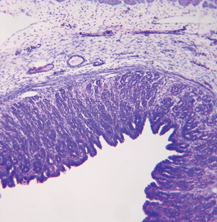
Your Stomach Is Not an Island
The control of the digestive process is one that involves an interaction between the nervous system, the endocrine system, the circulatory system, and the digestive system. Watch the following animation Hormones and Gastric Digestion about the regulation of digestion.
Pay particular attention to the positive feedback that reinforces the continued release of gastric secretions into the stomach. In addition, watch for the negative feedback that shuts off the release of these secretions when stomach digestion is complete.
For more information about feedback and homeostasis, check page 203 in the textbook.
pepsinogen: the inactive precursor to pepsin formed in the chief cells of the mucous membrane of the stomach and converted to pepsin by hydrochloric acid during digestion
pepsin: a digestive enzyme found in gastric juice that catalyzes the breakdown of protein to peptides
Hydrochloric acid and pepsinogen are secreted by the stomach as components of gastric juice. These secretions are signaled by nerve impulses and hormones when you taste, smell, touch, or think of food. Once in the stomach, pepsinogen is activated by the low pH levels created by the hydrochloric acid (HCl) and is converted to the active enzyme pepsin. Pepsin catalyzes the breakdown of proteins to smaller units called peptides.
 Try This
TR 2. Enroute from Gums to Bums
 Read
Read
© Sebastian Kaulitzki/shutterstock
Return to the Assignment to add the stomach, its functions, secretions, and the function of its secretions to your flow chart or diagram.
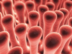
The small intestine is, in fact, much longer than the large intestine but because of its smaller diameter it has been labelled “small.” The small intestine has several structural elements that aid in its main function of completing the digestion of macromolecules and absorption of the subunits.
Several accessory organs provide digestive assistance through their secretions. The pancreas, liver, and gall bladder secrete enzymes that help to complete the digestion of macromolecules. In this way, the resulting subunits can be transported across cell membranes and delivered to the circulatory system for distribution to all body cells.
To help you complete the next Self-Check and Try This, you will need to read “Digesting and Absorbing Nutrients: The Small Intestine” on pages 222 to 228 of the textbook.
The small intestine has the largest role in the digestive system.
 Self-Check
SC 3. Do this Self-Check exercise about the small intestine.
Try This
TR 3. Enroute from Gums to Bums: The Small Intestine and Accessory Organs
Go to your Assignment to add the small intestine, its functions, its secretions, and the function of its secretions to your flow chart or diagram. Don’t forget to also include the pancreas, liver, duodenum, and gall bladder.
Read
The purpose of digestion is to break down macromolecules into useable components for growth and repair. Once digestion has finished, what does the body do next?
As you have come to see, no body system is independent of another. A complex network of circulatory vessels are highly concentrated in the villi of the small intestine.
As molecules become small enough to move across cell membranes, they are actively transported via transport proteins. Molecules, such as amino acids, move across the villi membrane where they then diffuse into the blood for circulatory transport to other body cells.
Glucose, a result of carbohydrate digestion, would be used by cells to generate ATP, which would power active transport. Diagrams on the bottom of pages 226 and 227 of the textbook illustrate the active transport of molecules across the cell membrane.
© Danilo Ascione/shutterstock
Read
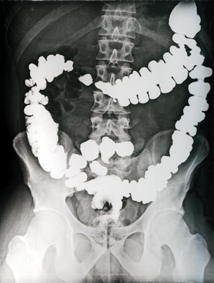
The colon is the main portion of the large intestine. Once chyme has entered the large intestine, there is no more digesting to be done. The main function of the intestine is to concentrate wastes and eliminate wastes.
By absorbing water and salts, the large intestine can reduce 500 mL of undigestible chyme to 150 mL of feces to be stored in the rectum and excreted.
Read page 231 of the textbook for a more detailed summary of the function of the large intestine.
Try This
TR 4. Enroute from Gums to Bums: The Large Intestine
Go to the Assignment to add the large intestine and its function to your flow chart or diagram.