Module 7 Intro
1. Module 7 Intro
1.18. Page 2
Module 7—The Digestive and Respiratory Systems
 Explore
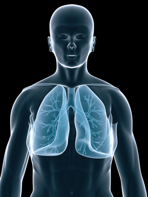
The Structure and Function of the Respiratory System

The main function of the respiratory system is to ensure that oxygen is brought to each cell in the body and that carbon dioxide leaves each cell and is removed from the body. Respiration is the general term that is used to describe this overall process.
With every breath you take, an invisible mixture of dust, fungi, bacteria, water, nitrogen, oxygen, carbon dioxide, and argon enters your respiratory tract. These substances are on a journey that will trap some of them in the cilia of the lung, sweep out others with your breath, and ultimately allow only one substance—oxygen—to pass into the bloodstream.
Oxygen from the atmosphere passes over warm, moist membranes in the nose and mouth known as the upper respiratory tract. Oxygen enters an ever-branching, ever-narrowing hair-lined network of passageways from the trachea to the bronchial branches.
Finally, oxygen reaches the more than a quarter-of-a-billion blind, grape-like sacs at the passageway’s end that is the alveoli in the lung. It is here that oxygen enters the circulatory system in exchange for carbon dioxide. The carbon dioxide then makes the reverse journey out of the body.
The next diagram shows the basic structure and function of the respiratory system. The Canadian Lung Association’s web page will help you get a better understanding of the labelled structures and has information on the process of respiration. By the end of this lesson, you should be able to give a clear and concise explanation of what each part of the respiratory system does.
To help you with the vocabulary associated with this lesson and as a support, a vocabulary review sheet is available to be downloaded.
 Try This
In Lesson 2 you conducted a virtual dissection of a pig’s digestion system. In this lesson you will use the same virtual dissection site to explore the pig’s respiratory system.
You may want to read pages 244 to 247 of your textbook before you perform a virtual pig dissection.
TR 1. Virtual Pig Dissection
When you are ready to begin the virtual lab, go to the online “Virtual Pig Dissection” and explore the “Respiratory System of the Virtual Pig.”
To find this virtual dissection, you will have to perform a web search using the keywords “virtual pig dissection Whitman.”
Once you have opened the dissection, read the instructions about necessary computer plug-ins. Proceed by clicking on “respiratory system” in the “STUDY GUIDES” box. Make sure that you follow all of the steps and click on all possible parts of the system indicated in the photos as “Things to Note.”
Once you have finished going through the entire dissection of the respiratory system, proceed to “QUIZZES” and click on “respiratory system.” There are several quizzes there, but in order to proceed to the next quiz you must obtain the correct answers. This is a good review of the respiratory system structures and functions. You may want to take screen captures of the dissection or the quizzes and save them to your course folder for review.
 Watch and Listen
Watch the following animation that shows the voyage of a molecule of oxygen through the structures that make up the respiratory passageways and the subsequent return of carbon dioxide to the atmosphere.
 Self-Check
SC 1. Answer this Self-Check exercise that has questions related to breathing.
© Sebastian Kaulitzki/shutterstock
There are two stages to breathing—inspiration and expiration. These are better known as breathing in and out. This movement of air into and out of the lungs is controlled by muscles that change the size of the pleural cavity (the thin, fluid-filled space between the two membranes that surround the lungs). This change in pleural cavity size affects pressure in the cavity relative to the external atmospheric pressure. Air naturally moves from a higher pressure to a lower pressure.
During inhalation, the pressure is greater in the atmosphere than it is in the pleural cavity because the size of the pleural cavity is increased, thereby decreasing its pressure.
 Read
Read
When exhalation occurs, the pressure in the pleural cavity is increased by decreasing its size. Read pages 249 and 250 in the textbook for more information.
Self-Check
SC 2. Complete this Self-Check exercise.
Try This
TR 2. The Mechanics of Breathing
The diaphragm and the rib muscles (internal and external intercostals) control the air pressure inside the lungs that causes air to move in and out of the lungs.
Go to your Lesson 3 Assignment where you will be asked to illustrate the processes of inhalation and exhalation.