Module 5
1. Module 5
1.4. Page 2
Module 5—Cell Division: The Processes of Mitosis and Meiosis
 Explore
 Read
Read
parent cell: a diploid somatic cell about to enter cell division
daughter cell: a cell that is the product of cell division
In mitosis, daughter cells are identical to the mother cell; in meiosis, they are not identical to the parent cell.
DNA: the genetic material found contained in the nucleus in eukaryotes (also in mitochondria and chloroplasts) and loose in the cytoplasm in prokaryotes, such as bacteria
histones: proteins found in chromosomes that provide scaffolding for DNA to twine around so that the DNA can fit within the confined space of the nucleus
chromatin: long fibres containing DNA, small amounts of RNA, and proteins
These fibres form chromosomes when they coil around histones.
centromere: a ‘button’ that holds the two identical sister chromatids together after the S phase of interphase and through mitosis until anaphase
To review some of the structures and the organization of cells that will be useful to you in this unit, read pages 546 to 547 and pages 550 to 552, up to “Chromosome Number,” in your textbook. Note the diagram on page 547 of the textbook, or play “Cellular Pursuit” on your own or with a friend as a way to review the different parts of a cell. From this reading or the game, create a glossary of terms needed in this unit. Include parent cell, daughter cell, DNA, chromosome, histones, chromatin, and centromere.
You will recall from earlier science courses that life does not spontaneously occur. Instead, life comes from existing life and is organized around small units called cells. The first part of the cell theory was proposed by two German biologists, Mathias Schleiden and Theodore Schwann. Based on their observations, they concluded that all plants and animals were made of cells. This conclusion has been extended to include all living things and, since their discoveries, no exceptions have been found. Particles such as viruses and prions are technically excluded from the list of “living things.”
© 2008 Jupiterimages Corporation
Types of Cell Division
The continuity of life from one cell to another is based on the reproduction of cells via cell division and occurs as part of the cell cycle. There are two major types of cell division: mitosis and meiosis.
Mitosis
© RTimages/shutterstock
Mitosis is simple cell replication. It begins with one parent cell and ends with two complete daughter cells, which are nearly identical to the original. If the cell being considered is a unicellular organism, then this kind of division is also a form of reproduction known as binary fission. Mitotic division in multicellular organisms is responsible for growth, development, and repair. To review why organisms must grow through cell division and not by increasing cell size, read page 550 of the textbook and consider “Figure 16.1” illustrating surface area to volume ratio.
binary fission: cell division in prokaryotes (bacteria); simple because there is only one circular chromosome so no spindle is needed
asexual reproduction: creation of a new organism without the input of cells from two separate organisms of opposite sexes; examples are binary fission, yeast and Hydra budding, and vegetative propagation of plants
cutting: type of vegetative propagation when a stem of a plant is cut off and produces roots, stems, leaves, and flowers; an asexual form of reproduction
variation: the existence of many combinations of genes/traits in a population; improves the probability that some members will survive if environmental conditions change; is high in sexual reproduction
mutation: a permanent change in a cell's genetic structure, often resulting in the expression of a new trait or feature in the affected organism; usually due to random errors occurring during DNA replication or protein synthesis, but can also be caused by chemical or physical mutagens
resistance: occurs when a drug removes susceptible bacteria or viruses from a population and leaves those variants (mutants) that are resistant to the drug
Rapid cell division ensures that the whole population becomes resistant quickly.
super bugs: bacteria that are immune to many antibiotics
Super bugs develop because of an overuse of antibiotics and antibacterials that have destroyed susceptible bacteria, leaving only those bacteria that are resistant to these drugs.
Some multicellular organisms can also reproduce through mitosis under special conditions. This is called asexual reproduction since it involves only one parent. Taking a cutting from a plant or cutting a flat worm in half are good examples. Some insects can also use this method to rapidly populate an area, like aphids that are born pregnant with more female aphids that will also be born pregnant!
The advantage of mitosis is in speed and energy costs. Only one cell is needed to start and from that many thousands of cells can result. This is why some bacterial infections can spread so rapidly. The disadvantage of this kind of replication is that daughter cells are genetically identical to the parent cell. Therefore, little or no variation exists in the population, and it may be susceptible to changes in the environment, such as the use of drugs to treat bacterial infection. However, some bacteria mutate very rapidly and develop a resistance to drugs. These are termed the super bugs, and they are a major concern in the medical community. In Unit D you will study the factors that determine whether or not a population is successful at surviving.
Courtesy of the National Center for Biotechnology Information/National Institutes of Health
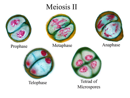
Meiosis
gametes: sex cells (sperm and egg); have half the normal chromosome number so they can participate in fertilization
fertilization: fusion of an egg and sperm (gametes) to produce a zygote; occurs in sexual reproduction only
sexual reproduction: creation of offspring through input of genetic material from two different organisms of opposite sexes (sperm from male and egg from female); increases variation
Meiosis is a more complex process. After a meiotic division, the resulting cells will have half the needed genetic information. These cells are called gametes. To complete replication, a gamete from one parent will need to unite with a gamete from another parent to restore the complete amount of genetic information. This is a process known as fertilization, which occurs during sexual reproduction.
While meiosis takes longer, involves more than one parent, and costs more energy to carry out, it does result in variation. By encouraging genetic variation in a population by using meiosis and fertilization, a species will be better suited to overcome change in the environment. Variation increases the chances for survival of a species and increases the chance that members of a population will reach reproductive age so that adaptive traits can be maintained in the population.
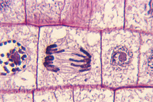
Genetic Material
The hereditary material carried by a cell is determined by the genetic code found on DNA (deoxyribonucleic acid). DNA is organized inside our body cells into segments called chromosomes. For most of a cell’s life, these segments of DNA are diffuse and cannot be observed under a microscope. However, when the cell undergoes division, the chromosomes condense and become visible. For more detail on the organization of chromosomes, read pages 551 and 552 in the textbook.
Every organism has a specific number of chromosomes in its cells. For example, human cells have 46 chromosomes, while dog cells have 78 chromosomes. This number must be maintained from generation to generation for normal function to occur. Chromosomes can be further organized into homologous chromosomes. Homologous chromosomes are roughly the same size and shape and contain the same type of genetic information.
somatic cell: the name given to any of the cells of a multicellular organism, including humans
The exception is those cells that form gametes, which are not somatic cells.
autosomes: the 22 homologous pairs seen in a karyotype; have nothing to do with gender
sex chromosomes: the last (twenty-third) pair of chromosomes seen in a karyotype that determines the gender of an organism
X and Y sex chromosomes are not homologous to each other in terms of shape, size, or genetic information.
X chromosome: the longer sex chromosome
Females are XX.
Y chromosome: the shorter sex chromosome; determines maleness; has much fewer genes on it than the X
Males are XY.
In each somatic cell of a human body there are 22 pairs of homologous chromosomes, known as autosomes, and one pair of sex chromosomes. The sex chromosomes determine the gender of the organism. A human with two X chromosomes is female, and a human with one X and one smaller Y chromosome is male. Read “Chromosome Number” on pages 552 to 553 in the textbook to learn more about chromosomes and how they are arranged in cells. You may choose to summarize this information as a glossary of terms. Include gene, locus, allele, diploid, haploid, and polyploid as terms in your glossary, as you will need to know their meaning as you work through this unit.
gene: the unit of hereditary information that can be passed on to offspring; includes the specific DNA sequence encoding or regulating the sequence of a protein, tRNA, or rRNA molecule; determines the expression of a trait
locus: a specific location on a chromosome
allele: a different form of the same gene occurring on homologous chromosomes
diploid: the term describing a cell that contains two pairs of every chromosome
haploid: the term describing a cell containing half the chromosomes that a diploid parent cell contains
This condition occurs in gametes, either in the egg or the sperm.
polyploid: the term describing a cell that contains more than two homologous chromosomes
Karyotype
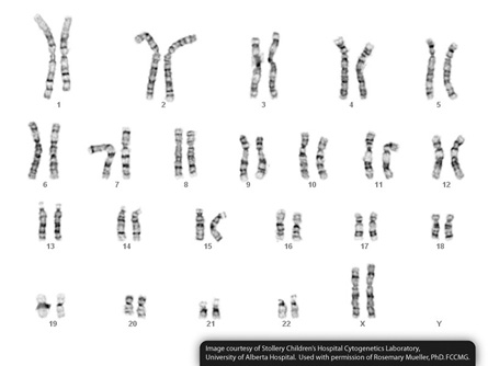
© Stollery Childrens Hospital
staining: a technique used in slide preparation to make the chromosomes of a dividing cell visible and dark
nondisjunction: an error in meiosis that results in non-separation of chromosomes; results in two chromosomes entering one gamete; produces gametes with an extra chromosome (n + 1), or gametes that are missing a chromosome (n – 1)
Down syndrome: typically characterized by some impairment of physical growth, unique physical features, and below average cognitive ability
If an n + 1 gamete that results from nondisjunction of a twenty-first chromosome is fertilized by a normal sperm, a Trisomy 21 (2n + 1) offspring is produced with Down syndrome.
Klinefelter syndrome: born with primary male sex characteristics but develops female secondary sex characteristics
When an XX egg due to nondisjunction is fertilized by a Y sperm, the offspring (XXY) has Klinefelter syndrome.
An individual’s chromosome set is known as their karyotype. The set, or number and arrangement of chromosomes, is very important for the result of regular life function. Scientists can make a picture of an individual's chromosome set by staining cells that are about to undergo cellular division because this is when the chromosomes are most dense. At this point, the chromosomes can be sorted into homologous pairs by their length, position of the centromere (a region of the homologous chromosome pair that appears pinched in), and banding pattern.
When sorted, scientists can determine the gender of an organism and whether or not an abnormal number of chromosomes is present. Certain major syndromes are a result of too many or too few chromosomes because chromosomes did not separate equally during cell division. This is termed nondisjunction. In humans, Down syndrome is a result of an extra chromosome 21 and Klinefelter syndrome is a result of an extra sex chromosome (XXY). Read “Examining Chromosomes: The Karyotype” on page 553 of your textbook.