Module 5
1. Module 5
1.9. Page 2
Module 5—Cell Division: The Processes of Mitosis and Meiosis
 Explore
For approximately 90% of a cell’s life cycle, it is not dividing. During this period of the life cycle, the cell performs normal vital activities, such as respiration, photosynthesis, protein synthesis, absorption, secretion, and excretion. Cell division, the remaining 10% of the life cycle, is a process involving a series of stages marking the beginning and end of cell division. Perhaps the most important process preparing for cell division is the replication of genetic material in the nucleus. This must be completed before a cell divides so that each daughter cell receives the same genetic complement as the parent cell. Once the replication is complete, the cell is ready to enter the process of division.
 Read
Read
Cell Cycle

Inquiry into Biology (Whitby, ON: McGraw-Hill Ryerson, 2007). 553, fig.16.5. Reproduced by permission.
Human bodies are made up of an amazing array of specialized cells that keep them healthy and able to maintain an internal balance in an environment that is constantly changing. During the daily struggle with the environment, cells must be replaced as they wear out. Exactly how fast cells are replaced is related to their role. Skin, which is constantly being scratched, rubbed, or cut, replaces itself very quickly. Muscle or nerve cells may remain healthy for most of a human's life and, therefore, may not need to divide to replace themselves. Some cells, such as red blood cells, which lack a nucleus and genetic material, or gametes, which only have one of each chromosome, will never reproduce.
The typical life cycle of a cell can be broken down into two main phases: interphase and M phase.
Interphase is the much longer phase, and is the phase where the cell will carry out its intended function in the body.
M phase, or mitosis phase, is shorter and moves the cell through a complex sequence of steps that are carried out in order to divide the cell’s genetic material equally. This phase ends with cytokinesis, which is the physical division of the cell into two daughter cells. Read pages 553 to 555 in your textbook to preview the stages of the cell cycle.
Interphase is further broken down into three phases: G1 phase, S phase, and G2 phase.
interphase: the longest period of the cell cycle when the cell is actively growing and metabolizing; consists of G1, S, and G2 phases; DNA is in loose, stringy chromatin form not visible under the microscope
M phase: mitosis and cytokinesis together
cytokinesis: the phase of the cell cycle after mitosis when the cytoplasm divides into two separate daughter cells
A cleavage furrow forms in animal cells; a division plate forms in plant cells.
G1 phase: the first part of interphase where the cell is actively growing and undergoing metabolism and protein synthesis
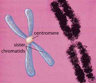
S phase: the second part of interphase where DNA replication occurs in preparation for upcoming mitosis; produces sister chromatids
G2 phase: the third part of interphase where the cell continues growing, metabolizing, and carrying out protein synthesis
Inquiry into Biology (Whitby, ON: McGraw-Hill Ryerson, 2007). 555, fig.16.6. Reproduced by permission.
During G1, or Growth 1, the cell is growing rapidly, producing proteins and carrying out its intended function. In the case of muscle or nerve cells, they may remain healthy and functioning at this stage for so long that they may be referred to as being “stuck” in G1 or, as it has more recently been referred to, in G0 phase. If, however, this is a regular cell, it will reach a point where it moves on to the S phase.
The G1 stage is critical if the cell is to divide properly later. In S phase, or synthesis phase, the cell will duplicate its DNA exactly. Each single chromosome makes a copy of itself and holds on to the copy. These doubled chromosomes do not contain any new genetic material. Rather, they are identical copies of each other. While together, they are known as sister chromatids, and are joined together by a regional structure called the centromere.
After the DNA has been successfully doubled, the cell will enter G2, or Growth 2. Here the cell continues to carry out its role in the body. Nearing the end of interphase, the cell will get ready for M phase by storing energy and building proteins and other structures needed for cell division.
During M phase, or mitosis phase, the cell will divide each doubled chromosome into two separate single chromosomes. This process will be covered in detail in the next lesson. At this point it should be clear to you that the cell goes through an orderly set of steps necessary to ensure that its genetic material is correctly divided into two complete and equal sets. Following this division, the cell will go through cytokinesis in order to physically divide the cell into two.
 Watch and Listen
Having learned the major divisions of the cell cycle, watch this animation, which brings each stage of the cell cycle together and illustrates how they would typically work in a eukaryotic cell. The animation also introduces you to three key checkpoints of the cell cycle: G1, G2, and M checkpoints. While you watch, consider why these checkpoints are important and why they occur when they do.
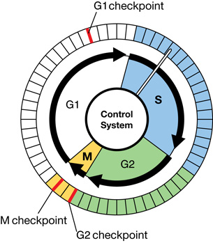
The animation introduced the checkpoints that regulate division within the cell. These checkpoints ensure that cells are growing and are capable of division.
There are three major checkpoints of the cell cycle. One is at the end of G1. Here, the cell evaluates if it is large enough and strong enough to continue with the division process. The second checkpoint is at the end of G2. This is a very important checkpoint for the cell. At this time, the cell must evaluate if it has properly duplicated all of its chromosomes. If it has not, it may attempt to carry on with the division, or it may simply self-destruct. The last major checkpoint occurs during M phase. At this checkpoint the cell evaluates whether the spindle apparatus has properly attached itself to each of the chromosomes, and whether the rest of the cell is ready for cytokinesis, or physical cell division. If something is wrong at this stage, the cell will often simply die.
spindle apparatus: a structure composed of spindle fibres; forms during prophase in mitosis to facilitate separation and movement of chromosomes in cell division
 Try This
To review the stages of the cell cycle and its checkpoints, do a web search using the search terms “educational games” and “control of the cell cycle.” In the activity that you will find, take on the role of Cell Division Supervisor. You will have a limited amount of energy to use for carrying the cell through all of the stages of cell division, as well as to check various cellular components to see if the cell is ready to move on. Try to keep the body healthy!
Read
Reasons and Limits for Cell Growth

There are many factors that may lead cells to reproduce. The most common reason is the need for replacement due to damage or age. Cells work very hard, and many simply become too brittle or build up toxins too fast to continue to function. These cells are broken down by the body, and their raw materials are reused.
Once the call for cell reproduction is given, how do cells know when to stop? Consider skin cells as a working example. Your skin is one of the most active areas on your body for cell reproduction. Skin cells use two common clues to determine when to start or stop reproduction. The first is density-dependent inhibition.
density-dependent inhibition: a property of normal cells that allows mitosis to occur only until cells touch each other
Density-dependent inhibition is lost in cancer cells; therefore, cells begin to form on top of one another, forming masses of cells called tumours.
For density dependent inhibition to work, cells respond to their neighbours. Skin cells will not divide if isolated. If a gap or a cut is created in a layer of skin cells, those that remain will automatically start to divide until the gap is covered. Once skin cells bump up against their neighbours, they stop dividing. The cut is healed.
Another clue that cues cell growth is anchorage dependence. Using skin cells again as the example, skin will grow naturally if anchored to a substratum of tissue. Conversely, they will not grow if simply free floating in a nutrient bath. For example, individual skin cells can’t be cultured in a nutrient bath, but skin cells anchored in tissue can be cultured to grow in a nutrient bath. These cultures are often used in treating severe burns with skin grafts.
anchorage dependence: a property of normal cells that only allows mitosis to occur when cells are attached to a substrate or surface, not floating freely
Anchorage dependence is lost in cancer cells, thereby allowing for metastasis to occur.
Cancer
cancer: rapid proliferation (cell division) of cells that occurs when mutations result in disruption of the normal timing of mitosis; characterized by loss of density-dependent inhibition, loss of anchorage dependence, dedifferentiation of cell function, rapid metabolism, and short cell cycle
The term cancer signifies a broad group of diseases characterized by a rapid, uncontrolled division of cells. Cancer cells appear to ignore all the regulations in place for cell growth. They do not wait or stop at any of the checkpoints. They do not appear to be density dependent, nor are they anchorage dependent. Cancer cells grow and reproduce constantly. They never enter the stage of the cell life cycle where specialized function occurs.
Watch and Listen
metastasis: the tendency of some cancer cells to break off from a primary tumour and move through the blood or lymphatic systems to other locations in the body where secondary tumours form; sometimes referred to as the “spreading” of cancer
Watch the following animations:
The first animation shows how a cancerous growth might occur. Another very dangerous characteristic of cancer is shown—its ability to spread. When cancer cells leave their original site, it is called metastasis. When cancer cells begin to spread in this fashion through the blood or lymphatic systems, it becomes much harder to get rid of them.
 Self-Check
Complete the multiple-choice questions about the cell cycle, and check your answers.
Watch and Listen
The video “Cell Cycle and Mitosis: Copying the DNA Blueprint” reviews the cell cycle and cancer. You may wish to view the video in order to review or reinforce these concepts and to find information related to the Lesson 2 assignment. Pay particular attention to the end of the video, as it goes over cancer treatments. Watch and listen for information related to the following questions:
-
radiation treatment: cancer treatment in which high-energy radiation from radioactive isotopes is directed at a cancerous tumour in an effort to destroy it without destroying surrounding normal tissueHow does radiation treatment help combat cancer?
- What are some of the considerations when using radiation treatment?
- What are some of the side effects of radiation treatment?