Module 8 Intro
1. Module 8 Intro
1.5. Page 3
Module 8—Circulation, Immunity, and Excretion
 Read
Read
Why Does the Heart Pump Rhythmically?
A healthy heart beats steadily and rhythmically at a rate of about 60 to 100 beats per minute when at rest (normal sinus rhythm). During strenuous exercise, the heart can increase the amount of blood it pumps up to four times the amount it pumps at rest, within only a matter of seconds. As a specialized organ, the heart depends on specialized muscle cells and fibres to pump in a coordinated, rhythmic fashion.
endocardium: the innermost layer of tissue that lines the chambers of the heart
This sheet of shiny, white tissue also lines the body’s blood vessels to help form a continuous lining through the circulatory system. This lining helps blood flow smoothly and prevents the formation of clots.
myocardial fibres: a specialized cardiac muscle that can contract as well as conduct electrical impulses; not found anywhere else in the body
Purkinje fibres: specialized fibres that transmit electrical impulses to the cardiac muscles in the heart to induce rhythmic muscle contraction
The pumping action (muscle contraction/relaxation) is coordinated by Purkinje fibres (or Purkyne tissue) located in the inner ventrical walls of the heart, just beneath the endocardium or lining of the heart chambers. These fibres are specialized myocardial fibres that conduct an electrical stimulus or impulse that enables the heart to contract in a coordinated fashion.
Read more about “The Beating Heart” on pages 272 to 274 of the textbook or watch this animation.

Inquiry into Biology (Whitby, ON: McGraw-Hill Ryerson, 2007), 274, fig. 8.7. Reproduced by permission.
Systemic vs Pulmonary Circulation
pulmonary: having to do with the lungs
systemic: a body system in general
It is important to distinguish between pulmonary circulation and systemic circulation. Pulmonary circulation occurs when the right side of the heart pumps deoxygenated blood to the lungs via the pulmonary arteries. Pulmonary veins bring oxygenated blood back to the left side of the heart to be distributed to the rest of the body through the arteries. In the pulmonary system, arteries carry deoxygenated blood and veins carry oxygenated blood.
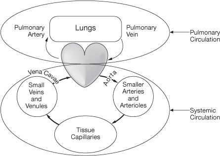
This is different from systemic circulation. Systemic circulation involves the circulation of blood to all other parts of the body aside from the lungs. This is the portion of the cardiovascular system that carries oxygenated blood away from the heart, to the body, and then returns deoxygenated blood back to the heart.
Read pages 268 to 270 in the textbook to obtain more details about the structure and function of the heart.