Module 8 Intro
1. Module 8 Intro
1.51. Lab
Module 8—Circulation, Immunity, and Excretion
 Lab: Urinalysis
You will now perform a modified urinalysis based on the investigation on page 320 of your textbook.
Problem
What physical and chemical tests can you use, and what data do they provide in the analysis of urine?
Procedure
Consider this scenario. A theft was committed in the washroom of a community building. Forensic specialists collected a urine sample at the scene of the crime. The police have four suspects in custody. Your task is to find out who committed the crime.
Open the Lesson 9 Assignment file, where there is a table to document the results of the tests and support the elimination of suspects. For each test, indicate the results for each individual in the chart. Once you have recorded the results for all of the tests, you should be able to identify the criminal.
You will start by interpreting the results of each test.
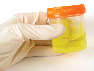
© Christina Richards/iStockphoto
Test 1—Colour, Odour, Clarity: Normal urine is a clear, straw-coloured liquid. Urine may be cloudy because it contains red or white blood cells, bacteria, or pus from a bladder or kidney infection. Normal urine has a slight odour. Foul-smelling urine is a common symptom of a urinary tract infection. A fruity odour is associated with diabetes mellitus. Use the following descriptions of the urine samples to fill in your chart located in your Lesson 9 Assignment.
Control: Urine is yellow. Smell is normal. Looks cloudy.
Crime suspect: Urine is yellow. Smell is fruity. Looks clear.
Suspect 1: Urine is yellow. Smell is fruity. Looks clear.
Suspect 2: Urine is yellow. Smell is normal. Looks clear.
Suspect 3: Urine is yellow. Smell is normal. Looks cloudy.
Suspect 4: Urine is yellow. Smell is fruity. Looks clear.
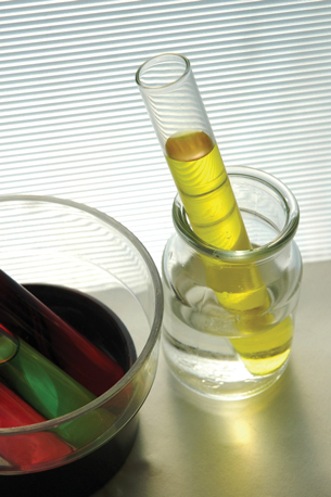
© mypokcik/shutterstock
Test 2—Protein: One sign of kidney damage is the presence of protein in urine. To test for protein, a portion of each sample is placed in a hot water bath. If the sample becomes cloudy or is cloudier than the original sample, it contains protein.
- After a few minutes, the test tube from the hot water bath is removed and compared to the original sample.
- If the heated sample is cloudier, it contains protein. The test is positive.
Use the following descriptions of the urine samples to fill in your chart located in your Lesson 9 Assignment.
Control: positive
Crime Suspect: negative
Suspect 1: negative
Suspect 2: negative
Suspect 3: positive
Suspect 4: negative
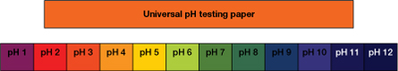
Test 3—pH: The wide range of pH values (pH 4.7 to 8.5) makes this the least useful parameter for a diagnosis of kidney disorders. Kidney stones are less likely to form and some antibiotics are more effective in alkaline urine. There may be times when acidic urine may help prevent some kinds of kidney stones. Bacterial infections also increase alkalinity, producing a urine pH in the higher 7–8 range.
A clean medicine dropper is used to place a drop of each sample of urine on small pieces of universal indicator (pH) paper. They are left for about 30 seconds. The pH paper can turn several different shades, depending on pH. pH can be determined by comparing the indicator to a colour chart.
Use the following descriptions of the urine samples to fill in the chart located in your Lesson 9 Assignment. To determine pH, use this indicator colour chart.
Control: yellow green
Crime Suspect: yellow/green
Suspect 1: orange
Suspect 2: green/blue
Suspect 3: green
Suspect 4: yellow/green
© Norman Chan/shutterstock
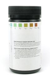
Test 4—Glucose: One sign that a person has diabetes mellitus is the presence of glucose in the urine.
- To test for glucose, a glucose test strip is dipped into each sample of unheated urine and immediately taken out.
- The strip is compared to a colour chart. The chart shows a colour gradient from light blue to dark brown. Intermediate colours are greens and browns. Blue indicates no sugar, while dark brown indicates high glucose levels. Intermediate colours indicate the presence of glucose in varying concentrations.
Use the following descriptions of the urine samples to fill in your chart located in your Lesson 9 Assignment.
Control: dark brown
Crime Suspect: dark brown
Suspect 1: light blue
Suspect 2: green
Suspect 3: light blue
Suspect 4: dark brown
From the results of the urine analysis, you should be able to tell which suspect committed the crime, or at the very least left his or her urine at the crime scene.
Complete the Analysis questions in the Lesson 9 Assignment.