Module 1
1. Module 1
1.27. Page 2
Module 1—The Nervous System
 Explore
The Structures of the Eye
 Read
Read
refract: to bend light as it passes through a substance with a different desnsity
retina: the innermost layer of the eye that contains the photoreceptors
optic nerve: a collection of sensory neurons that carries sensory information from the photoreceptors to the brain
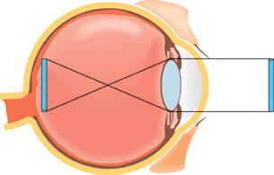
The eye is an amazing organ! It contains many structures that must all be functioning properly in order for you to see. Several of these structures refract, or bend, light and focus it on the different photoreceptors that make up the retina. Once the photoreceptors are stimulated by light energy, they convert it into an electrochemical nerve impulse. Sensory neurons in the optic nerve communicate this nerve impulse to interneurons in the occipital lobe of the brain, where visual information is then processed. To understand the parts of the eye and how they contribute to vision, read from page 410 to the heading “Conditions Affecting the Cornea and Lens” on page 412 of the textbook.
As you read, keep track of the vision disorders and their respective symptoms. You will need this information for an upcoming assignment in which you will be required to develop a summary chart of eight vision disorders. For each disorder, you will include information about the structure affected, the symptoms of the disorder, the physiology of the disorder, and technologies that can be used to detect or treat the disorder.
It is always an excellent idea to support your understanding with a diagram. You may choose to include diagrams in the work you will store in your course folder. If you choose to include diagrams, be sure to include brief descriptions of the functions of any structures and brief summaries of the steps of different pathways.
 Try This
There are three Try This activities in this lesson that can be used to verify your understanding of the parts of the eye and their functions as well as vision issues related to the dysfunction of one or more of these structures. There is a choice of two possible activities for each one. You may choose to do all of the Try This activities for a complete review of the concepts you have been studying.
TR 1. Choose one of the following activities.
Complete the eye function drag-and-drop activity to review the structures and functions of the eye.
OR
Watch the “Bio Review: Eye Structure and Function” section of “The Eye: Vision and Perception: A Whole World to See” that deals with the structures of the eye. You may be required to enter a username and password in order to access this video. Contact your teacher for this information.
After watching the video, make a diagram of the eye that includes summary notes about the function of each part of the eye. Store this work in your course folder for review purposes.
Accommodation
ciliary muscle: a ring of muscle behind the iris that is attached to the lens by suspensory ligaments and is involved in changing the shape of the lens
accommodation: the process of changing the shape of the lens from round and fat to thin and flat, and vice versa, so that light can be focused on the retina to accommodate vision of objects near and far away
The ciliary muscle and suspensory ligaments are excellent examples of how the sympathetic and parasympathetic nervous systems work together to establish homeostasis in vision. By changing the shape of the lens, light rays are bent differently, allowing you to focus on the words of this sentence or on the words on a poster across the room. This process is called accommodation.
Try This
TR 2. Choose one of the following activities.
Put the following lists in the correct order. You may wish to look at “Figure 12.10” on page 412 of the textbook to help you figure out the correct order of events. This activity is similar to the numerical-response questions that you will complete on the Diploma Exam. Note that the number assigned to the event does not imply the correct order. In this type of numerical response, you take the assigned number of the event and place that number in the correct order or sequence in the response area.
- Sympathetic Nervous System
- tension pulls on lens
- focus on far objects
- lens flattens
- tension on suspensory ligament
- ciliary muscle relaxes
Place the numbered events in the order that illustrates the correct order or sequence in which these events occur.
___, ___, ___, ___, ___
- Parasympathetic Nervous System
- ciliary muscle contracts
- tension on suspensory ligament relaxes
- less pull on lens
- lens bulges
- focus on near objects
Place the numbered list in the correct order.
___, ___, ___, ___, ___
OR
Review the accommodation reflex by watching “Bio Review: Accomodation Reflex” segment of “The Eye: Vision and Perception: A Whole World to See.”
You may be required to enter a username and password to be able to view the video. Contact your teacher for this information.
Try This Answers
TR 2.
- The correct numerical sequence of events is 5, 4, 1, 3, 2.
- The correct numerical sequence of events is 1, 4, 3, 2, 5.
The Lens
Read
The flexibility of the lenses decreases as people age. As a result, many people must wear reading glasses to focus the details of nearby objects. Hyperopia is the term used to describe the ability to see distant objects clearly and the inability to focus on objects that are close. You likely know the more common term for this condition—farsightedness. Myopia is the condition of being able to clearly see objects that are close but not being able to focus on objects that are farther away. The common term for myopia is nearsightedness.
To review what enables the lens to change its shape in order to successfully focus an image, you may wish to review pages 412 and 413 in the textbook. When you have finished the reading, make summary notes about the concepts you learned. You may also wish to draw a labelled diagram. Store your information in your course folder for review purposes.
Cataracts are another condition that affects the lens. Astigmatism is a condition that affects the cornea. Both these conditions distort vision. Remember to add them to your table of disorders. You may choose to research these conditions and add the information about these disorders to your table now. At the end of this lesson, you will submit your table to your teacher for assessment.
To develop your mastery of the concepts you have been reading about, do questions 1, 2, 3, and 5 on page 418 of your textbook. You may discuss your responses with your teacher if you wish.
hyperopia: farsightedness, or the inability to focus objects that are close, caused by an eyeball that is too short, which causes light to be focused behind the retina
myopia: nearsightedness, or the inability to focus objects that are far away, caused by an eyeball that is elongated, which causes light to be focused in front of retina rather than directly on it
cataract: a cloudy or grey-white area on the lens caused by deterioration of the protein composing the lens
Cataracts prevent the passing of light to the photoreceptors of the retina.
astigmatism: an uneven curvature of the cornea or lens, resulting in uneven focusing, which results in poor vision
cornea: a transparent portion of the sclera (located at the front of the eye) that allows light to enter the eye and, in the process, refracts or bends the light rays so that they can be focused on the retina
The Retina and Supporting Structures

© Tim Mainiero/shutterstock
The inner layer of the eye consists of the delicate retina, which contains two different types of photoreceptors called rods and cones. Directly in line with the middle of the lens is an area of the retina called the fovea centralis, or fovea. This small, depressed area is where the cones are most highly concentrated. Cones are the photoreceptors that are sensitive to different colours, whereas rods are sensitive to light intensity. Rods are more spread out on the periphery of the retina. The neural fibres from the retina form the optic nerve, which is the sensory pathway between the eye and the brain. The spot where the fibres come together is the blind spot. This area is called the blind spot because it lacks receptor cells. Therefore, any light or image falling on this area goes undetected.
Organisms that see well at night, like deer and cats, have a reflective layer, similar to spray paint, on the surface of the choroid layer. This structure is called the tapetum. The tapetum increases the animal’s sensitivity to low levels of light and helps these animals see at night. This layer also causes the eyes of these animals to appear iridescent or reflective in the dark.
In front of the lens is the anterior chamber, which is filled with aqueous humour. This transparent, watery fluid is produced by the ciliary body. If the aqueous humour is not successfully drained from the anterior chamber, its buildup will eventually result in the eye disorder called glaucoma. Behind the lens is the posterior chamber filled with vitreous humour—a clear to amber-coloured gel-like fluid. Both the aqueous and vitreous humours will aid the refraction of light to a small degree.
rods: one of two types of photoreceptors in the retina of the eye that is sensitive to light intensity and detect movement
Rods do not distinguish colour.
cones: one of two types of photoreceptors in the retina of the eye that is sensitive to different wavelengths of light and are, thus, responsible for distinguishing colour
There are three types of cones: one sensitive to red light, one sensitive to blue light, and one sensitive to green light. The cones are responsible for acute vision, or distinguishing detail.
fovea centralis: an area of the retina that is located directly behind the centre of the lens and has a very high concentration of cones, which makes this part of the eye responsible for great visual acuity
blind spot: the area at the back of the eyeball that is deficient in rods and cones; the area where the sensory fibres come together to form the optic nerve
choroid: the middle layer of the eyeball that lies between the sclera and retina and is highly vascular and heavily pigmented
The choroid absorbs stray light rays not detected by the photoreceptors of the retina.
tapetum: a layer in the choroid that increases the absorption of light to stimulate photoreceptors in dim conditions
anterior chamber: the space in front of the iris and behind the cornea that is filled with aqueous humour
aqueous humour: a clear, watery fluid in the anterior chamber of the eye that maintains the shape of the cornea and provides oxygen and nutrients for the surrounding cells, including those of the lens and the cornea
glaucoma: a disorder caused by the malfunction of ducts that drain excess aqueous humour from the anterior chamber
The resulting pressure created by excess aqueous humour ruptures delicate blood vessels in the eye and causes deterioration of cells in the eye due to lack of nutrients. This can result in blindness if left untreated.
vitreous humour: the transparent, amber-coloured, jelly-like fluid in the posterior chamber of the eye that helps to maintain the shape of the eyeball
Try This
TR 3. Choose one of the following activities. Whichever one you choose, be sure to take summary notes and then save them in your course folder.
Review the parts and functions of the eye by reading pages 414 and 415 in your textbook.
OR
Watch the following segments of “The Eye: Vision and Perception: A Whole World to See”
- “Bio Probe: Cow Eye Dissection”
- “Bio Review: Eye Structure and Function”
You may be required to enter a username and password to be able to view the video. Contact your teacher for this information.