Module 1
1. Module 1
1.30. Page 5
Module 1—The Nervous System
Rods and Cones Initiate Nerve Impulses

Inquiry into Biology (Whitby, ON: McGraw-Hill Ryerson, 2007), 415, fig. 12.15. Reproduced by permission.
Rods and cones contain pigments called photopsins, which absorb light and start chemical changes. Cones contain three different pigments called iodopsins. Rods have only one type of pigment called rhodopsin. When rhodopsin absorbs light, it is bleached and breaks down into opsin, a colourless protein, and a derivative of Vitamin A called retinal. This chemical change in the rods initiates a nerve impulse in the bipolar cells, which transfers the impulse to the ganglions. The ganglions, which make up the optic nerve, then transmit the impulse to the occipital lobe of the cerebrum to be processed.
Your eye then restores opsin and retinal to rhodopsin, but this takes energy. As you will recall from Biology 20, this energy is in the form of ATP. This reverse reaction also requires Vitamin A. Knowing this, can you explain why your eyes get strained in poor lighting, why night blindness is caused by Vitamin A deficiency, and why you should eat your carrots?
iodopsin: the general name of any of the three visual pigments found in cone cells that is stimulated by light to initiate a nerve impulse
rhodopsin: a visual pigment found in rod cells that is decomposed by light into opsin and retinal
The change from rhodopsin to opsin and retinal initiates a nerve impulse.
opsin: a protein that is the result of the decomposition of rhodopsin
retinal: a derivative of Vitamin A (retinol) that is the result of the decomposition of rhodopsin and is instrumental in initiating a nerve impulse
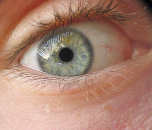
© Gilmanshin/shutterstock
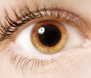
© Johanna Goodyear/shutterstock
Light and Dark Adaptation
Rhodopsin is extremely sensitive to light. Even starlight causes some molecules of rhodopsin to become bleached. In high-intensity light, rhodopsin is broken down almost as fast as it is remade. In such a situation, the rods become nonfunctional and the cones take over completely. For example, think about what happens when the lights are turned on after a movie. You are momentarily “blinded,” and all you see is white light. Your pupils constrict as a reflex to protect your retina from the bright light. Within a minute or so, the cones become sufficiently stimulated to take over once again. Over the next few minutes, visual acuity and colour vision improve. On the other hand, adaptation to low light occurs when you go from a well-lit area into a dark one. At first you see nothing but velvety darkness because your cones have stopped functioning due to lack of light, and your rods are not functioning because they have been bleached out by the bright light. Once in the dark, rhodopsin is remade and accumulates so that low-intensity light can again stimulate the rods. During adaptation to both light and dark, reflexive changes occur in the pupil.
The Pathway to the Brain
 Try This
TR 5. Did you know that each eye sees thing differently? Each eye has its own area of “blindness,” the blind spot. To find your blind spot, try the activity shown in “Figure 12.17” on page 416 of your textbook.
The information you receive from each eye is communicated through the optic nerve to the brain. Your brain processes this information into one image without gaps or blind spots. The optic nerves enter the optic chiasm on the ventral, front of the brain (see Lesson 2). In the X-shaped optic chiasma, some axons from each eye cross over to the opposite side of the brain, while others continue to their own side of the brain. The crossing over of about half of the sensory fibres ensures that each half of the occipital lobe receives the same image or part of the visual field viewed by each eye. However, keep in mind that each eye will see the image from a slightly different angle. Study the illustration provided below very carefully in order to better understand visual interpretation by the brain.
depth perception: the ability to see in three dimensions
Depth perception is possible due to the “fusing” or superimposing of the slightly different images from the two eyes by the visual cortex. This creates a three-dimensional image. Thus, by working together, the eyes provide slightly different angles of view that allow the brain to estimate distance.
The image that was reversed and inverted in the lens is at this point interpreted correctly.
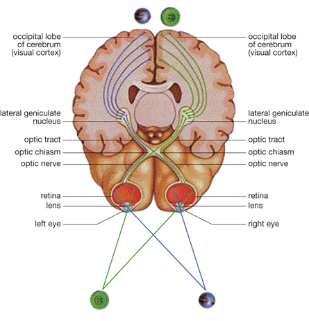
Inquiry into Biology (Whitby, ON: McGraw-Hill Ryerson, 2007), 416, fig. 12.18. Reproduced by permission.