Module 1
1. Module 1
1.53. Page 2
Module 1—The Nervous System
 Explore
Anatomy of the Synaptic Gap
 Read
Read
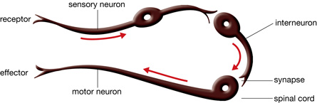
Your retinal receptor cells registered the new girl at school, the olfactory receptors detected the smell of her perfume, and the pressure receptors were activated by the contact of your handshake. The receptor cells converted this information to a nerve impulse, and information was on its way through a sensory neuron. But it came to a screeching halt at the end of the sensory neuron. Where does it go now? How does communication get to the next neuron? In the world of neurons, there’s a big gap to jump to the next neuron. To understand where the transmission goes next and the structures involved in the leap across the gap, study “Figure 11.18” on page 379 of your textbook.
synaptic knob: the tiny enlarged ending on an axon terminal
synaptic vesicle: a tiny membranous sac that, in this case, contains neurotransmitters
neurotransmitter: a chemical messenger released from the synaptic knob of a neuron at a synapse that diffuses across the synaptic cleft, binds to specially shaped protein receptors on the postsynaptic membrane, and stimulates the postsynaptic neuron
presynaptic membrane: the surface membrane surrounding the synaptic knob and facing the synaptic cleft
presynaptic neuron: the sending neuron
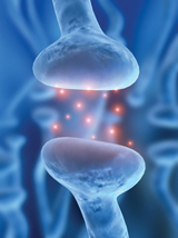
synaptic cleft: a tiny space separating the synaptic knob of a transmitting neuron from a receiving neuron or effector cell
postsynaptic neuron: the receiving neuron
postsynaptic membrane: the surface membrane on the dendrites of a receiving neuron across the synaptic cleft; contains receptor sites for neurotransmitters
Note the role of “shape” in controlling biological processes.
The synaptic knob is surrounded by the presynaptic membrane. The neuron that ends in the synaptic knob is called the presynaptic neuron. The synaptic cleft is the space between the presynaptic neuron and the next neuron, called the postsynaptic neuron. Remember that the receiving parts of the neuron are called the dendrites. Notice in the diagram on page 379 that the membrane surrounding the dendrite is called the postsynaptic membrane. Find the sodium ion channels in the postsynaptic membrane. Notice how these channels are only open when the neurotransmitter fits into a receptor. Note the role of shape in controlling biological processes.
© iDesign/shutterstock
© iDesign/shutterstock

© David Hughes/shutterstock
© Sebastian Kaulitzki/shutterstock
Crossing the Divide
Have you ever stood on a rock in a stream and wondered whether you could leap to the next rock? You think you can do it—you’re strong and co-ordinated—but if you fall, you could get very wet. To understand how a nerve transmission takes the “big leap” across the synaptic gap, study “Figure 11.18” on page 379 of the textbook and read "Signal Transmission Across a Synapse" on pages 378 to 380. Then follow up by looking at the animation showing synaptic transmission.
The diagram and readings illustrate that when the nerve impulse arrives at the synapse, it stimulates several reactions at the end of the axon. There is movement of the synaptic vesicles toward the presynaptic membrane, and then the vesicles fuse with the membrane. A neurotransmitter is released into the synaptic cleft, and it quickly diffuses across the synapse. Neurotransmitter molecules lock into receptor molecules on the postsynaptic membrane, causing the sodium gates to open and sodium ions from the synaptic cleft to rush into the postsynaptic neuron. You have also already learned that an inflow of sodium ions causes depolarization, and the start of an action potential. This now happens in the postsynaptic neuron (dendrite), resulting in a wave of depolarization through the postsynaptic neuron. The presynaptic and postsynaptic neurons have communicated a message across the gap via a type of chemical messenger called a neurotransmitter.
Summarize these events for your course folder as notes, a labelled diagram, a flow chart, or any format that you prefer.
 Watch and Listen
To visually explore synaptic transmission, you should choose to do one of the following:
- Watch the following segment of "Nerve Impulse Conduction: Dentists Calm Your Nerves." You may be required to enter a username and password to access the videos. Contact your teacher for this information.
- “Impulse Travel”
- “Impulse Travel”
- Watch the following segments of "Reflexes and Synaptic Transmission: Getting the Message Across."
- “Synaptic Transmission”
- “Bio Review: Synaptic Transmission”
As you watch any of the videos, answer the following questions. These questions will be an excellent reference for studying.
- Outline the reward pathway.
- What is a synapse?
- What happens at a synapse during synaptic transmission?
- What is a synaptic cleft? Is a synaptic cleft different from a synapse?
- What is a synaptic vesicle?
- What is the neurotransmitter in the reward pathway?
- Where are dopamine receptors located?
- What does the arriving nerve impulse do?
- What happens to the released neurotransmitter?
- Why is there a sucking sound in the video?
- What happens to the dopamine?
- How is the nerve impulse started in the second neuron?
- Identify two things that stop nerve impulse transmission.
- Does one neuron always synapse only with one other neuron? Suggest where this might be true.